Monday 05 May 2025
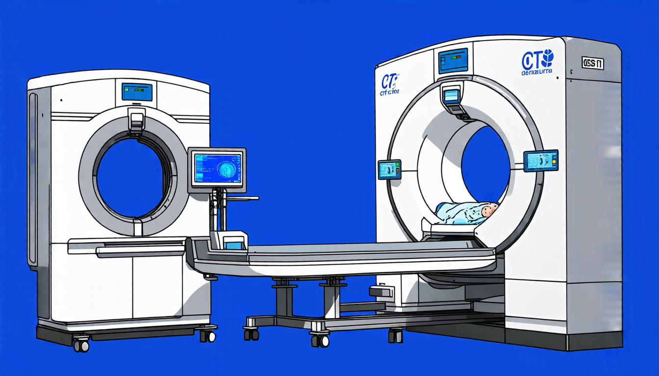
A team of researchers has made a significant breakthrough in developing a new technique for reconstructing dual-energy cone-beam computed tomography (DECBCT) images using only two orthogonal X-ray projections. This approach, known as 2V-DECBCT, has the potential to revolutionize medical imaging by enabling fast and low-dose DE volumetric imaging with high spectral fidelity and structural accuracy.
Conventional single-energy CBCT has limitations in image artifacts and poor soft-tissue contrast stemming from scattered radiation and beam-hardening. Dual-energy CBCT (DECBCT) has been proposed as a solution to overcome these limitations, but it requires full-trajectory scanning, which is time-consuming and susceptible to patient motion artifacts.
The researchers have developed a pair of diffusion models that operate in parallel across both the projection and image domains, corresponding to each energy channel. Through a cycle-domain training mechanism, physics consistency between projections and reconstructions is enforced at every diffusion step. In addition, the model incorporates a spectral-consistency constraint into the training process, which encourages the two energy-specific networks to preserve the expected differential contrast across energy channels.
The results of this study demonstrate the technical feasibility and potential of 2V-DECBCT reconstruction. The reconstructed images show improved structural accuracy and reduced streaking artifacts compared to conventional single-energy CBCT reconstructions. Energy subtraction maps computed from the reconstructed DECBCT volumes also exhibit high spatial agreement with reference maps generated from full-view dual-energy data.
This new technique has significant implications for medical imaging, particularly in image-guided radiation therapy where precise localization and rapid online adaptation are critical. The ability to reconstruct DECBCT images using only two orthogonal X-ray projections could enable fast and low-dose imaging, reducing patient exposure and improving the overall quality of care.
The researchers plan to further evaluate their technique using simulated patient datasets derived from CT scans of real patients, as well as physical phantom studies acquired on real scanners. This will enable them to assess generalizability, identify failure modes under unseen anatomies, and benchmark clinical applicability in realistic scenarios.
Overall, this innovative approach has the potential to transform medical imaging by providing a fast, low-dose, and high-contrast DECBCT imaging solution that can be used in a wide range of clinical applications.
Cite this article: “Fast and Low-Dose Dual-Energy Cone-Beam Computed Tomography Imaging”, The Science Archive, 2025.
Medical Imaging, Dual-Energy Ct, Cone-Beam Ct, X-Ray Projections, Reconstruction Algorithm, Diffusion Models, Physics Consistency, Spectral Fidelity, Image Artifacts, Low-Dose Imaging