Sunday 14 September 2025
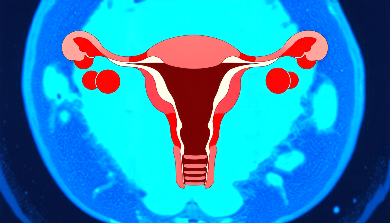
A team of researchers has made a significant breakthrough in developing an automated system for segmenting uterine myomas on MRI scans, a crucial step towards more accurate and efficient diagnosis and treatment of this common condition.
Uterine fibroids are the most common benign tumours of the female reproductive system, affecting approximately 70% of women of childbearing age. While they can cause symptoms such as abnormal bleeding, infertility, pelvic pain, and pressure-related discomfort, treatment decisions are largely influenced by the size, number, and anatomical location of the fibroids.
Magnetic Resonance Imaging (MRI) is a non-invasive imaging modality commonly used in diagnosis, but segmenting uterine fibroids requires a precise assessment of both the uterus and fibroids on MRI scans. This process is currently labor-intensive and time-consuming, and subject to variability due to intra- and inter-expert differences at both pre- and post-treatment stages.
To address this challenge, researchers have developed a deep learning-based automated segmentation method for uterine myomas using a publicly available MRI dataset. The proposed method uses a U-Net-based encoder-decoder architecture with five resolution levels, which integrates convolutional blocks with instance normalization and leaky ReLU activation.
The method was trained on 246 patients’ data and tested on 54 scans, achieving a mean Dice score of 0.70 for myoma segmentation, indicating high accuracy in detecting and delineating fibroids across diverse cases. The method also performed impressively across other classes, including the uterine wall (0.86), uterine cavity (0.79), and nabothian cyst (0.68).
The results demonstrate that the proposed automated system can reliably segment uterine myomas along with surrounding structures, offering a robust baseline for future research. This approach holds promise for reducing the clinical burden associated with manual segmentation and enabling more standardized assessment of fibroid characteristics.
This breakthrough has significant implications for women’s reproductive health, as accurate diagnosis and treatment of uterine fibroids are crucial steps towards alleviating symptoms and improving quality of life. The development of automated segmentation methods will facilitate more efficient and effective diagnosis and treatment strategies, ultimately benefiting patients and healthcare providers alike.
Cite this article: “Automated Uterine Myoma Segmentation on MRI Scans”, The Science Archive, 2025.
Uterine Fibroids, Mri Scans, Deep Learning, Automated Segmentation, U-Net Architecture, Instance Normalization, Leaky Relu Activation, Dice Score, Myoma Detection, Women’S Reproductive Health


