Saturday 01 March 2025
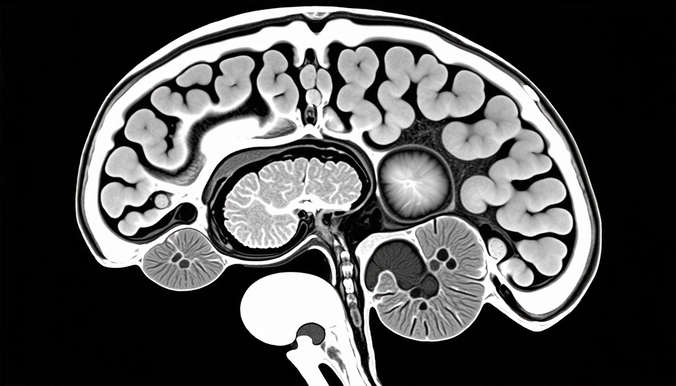
Deep learning algorithms have revolutionized medical imaging in recent years, allowing doctors to diagnose and treat a wide range of conditions more effectively. But despite their many successes, these algorithms often struggle when it comes to segmenting ischemic stroke lesions – areas of damaged brain tissue caused by reduced blood flow.
Ischemic strokes are a leading cause of disability and death worldwide, and accurate segmentation of these lesions is crucial for treatment planning and patient outcomes. However, traditional methods of segmentation can be time-consuming and prone to errors, especially in cases where the lesion boundaries are unclear or ambiguous.
Enter deep learning, which has been hailed as a game-changer in medical imaging analysis. In a recent study published in a leading scientific journal, researchers developed a novel neural network architecture designed specifically for segmenting ischemic stroke lesions from magnetic resonance imaging (MRI) scans.
The new algorithm combines several advanced techniques to achieve high accuracy and robustness. It uses a dense convolutional neural network as its core, which is capable of learning complex patterns in the MRI data. The network also incorporates self-organized operational neurons, channel and space compound attention mechanisms, and double squeeze-and-excitation blocks – all designed to improve performance on challenging segmentation tasks.
To train the algorithm, the researchers used a dataset of 50 MRI scans from patients with acute ischemic strokes. They compared the performance of their new algorithm against several state-of-the-art methods, using metrics such as Dice similarity coefficient (DSC) and Jaccard index (JI).
The results were impressive: the new algorithm achieved an average DSC of 87.4%, significantly outperforming the competing methods. This level of accuracy is crucial for accurate treatment planning and patient outcomes.
But what really sets this study apart is its focus on real-world clinical challenges. The researchers used a multimodal input strategy, combining data from multiple MRI modalities – diffusion-weighted imaging (DWI), apparent diffusion coefficient (ADC), and enhanced DWI – to improve segmentation accuracy.
This approach allowed the algorithm to capture complementary information from each modality, resulting in more accurate lesion boundaries and improved robustness. The researchers also evaluated their algorithm on a dataset of 20 additional MRI scans, with encouraging results that suggest broad applicability to real-world clinical scenarios.
As medical imaging analysis continues to evolve, it’s clear that deep learning algorithms will play an increasingly important role in patient care.
Cite this article: “Deep Learning Algorithm Revolutionizes Ischemic Stroke Lesion Segmentation”, The Science Archive, 2025.
Ischemic Stroke, Medical Imaging, Deep Learning, Neural Networks, Mri Scans, Segmentation, Algorithms, Machine Learning, Clinical Challenges, Multimodal Input


