Thursday 06 March 2025
Cardiac imaging just got a major boost with the development of a new technique that can accurately map the heart’s T1 values at ultra-high magnetic fields. This breakthrough has significant implications for our understanding and diagnosis of cardiovascular diseases.
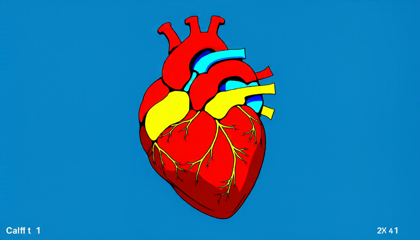
The heart is a complex organ, and its function depends on the precise alignment of its various components. One key aspect of this alignment is the relaxation time of the heart’s tissues – known as T1 values. These values can reveal important information about the heart’s health, such as the presence of scar tissue or inflammation.
However, measuring T1 values at ultra-high magnetic fields has proven to be a challenge. The high energies involved make it difficult to accurately capture the signals from the heart, leading to inconsistent results and limited resolution.
Enter the new technique, known as mIR-rt (multi-inversion recovery with real-time sampling). This method uses advanced pulse sequences and real-time acquisition to generate highly accurate T1 maps of the heart. By employing multiple inversion pulses and carefully timing the acquisitions, researchers were able to overcome the limitations of previous techniques and achieve unprecedented resolution.
The results are impressive. The new technique has been tested on phantom models and human subjects, yielding T1 values that closely match those obtained using reference methods. Moreover, the mIR-rt sequence is capable of producing detailed maps of the heart’s T1 values, revealing subtle variations in tissue relaxation times that could not be detected previously.
The implications of this breakthrough are significant. By allowing for more accurate diagnosis and monitoring of cardiovascular diseases, mIR-rt has the potential to revolutionize our understanding of the heart and its many complexities. This new technique could also enable researchers to study the effects of disease on the heart’s tissues in unprecedented detail, paving the way for the development of novel treatments.
The next step is to refine the technique and apply it to a wider range of clinical scenarios. Researchers are already exploring ways to optimize the mIR-rt sequence for use in real-world settings, where the challenges of imaging the heart can be even greater. With its potential to improve diagnosis and treatment outcomes, this new technique has the power to transform our understanding of cardiovascular health – and ultimately, save lives.
Cite this article: “Groundbreaking Technique Maps Hearts T1 Values at Ultra-High Magnetic Fields”, The Science Archive, 2025.
Cardiac Imaging, Magnetic Resonance Imaging, T1 Values, Cardiovascular Diseases, Heart Mapping, Multi-Inversion Recovery, Real-Time Sampling, Advanced Pulse Sequences, Phantom Models, Human Subjects