

Friday 14 March 2025
Medical imaging has long been a crucial tool in diagnosing and treating diseases, but it’s often a painstaking process for radiologists. Chest X-rays are particularly challenging, as they can be difficult to interpret and require extensive training to identify abnormalities. A new study is working to change that by developing an artificial intelligence system capable of accurately detecting thoracic diseases from chest X-ray images.
The researchers employed a deep learning approach, using convolutional neural networks (CNNs) to analyze the images. They trained their models on a massive dataset of over 112,000 frontal-view chest X-rays labeled with 14 different thoracic disease conditions. The CNNs were able to learn complex patterns in the images, such as shapes and textures, allowing them to identify signs of disease.
One of the key challenges in developing an AI system for medical imaging is addressing class imbalance. In this case, some diseases are much more common than others, making it difficult for the model to learn from the less frequent conditions. The researchers addressed this issue by using a loss function called focal loss, which prioritizes learning from the harder-to-classify examples.
The results were impressive: the AI system was able to achieve high accuracy in detecting a range of thoracic diseases, including atelectasis and effusion. Furthermore, the models were able to focus on specific regions of the images that were most relevant to the diagnosis, providing valuable insights for radiologists.
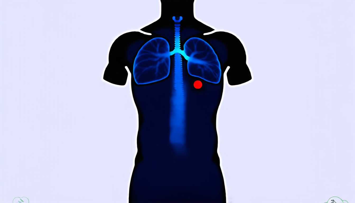
The researchers also employed a technique called Grad-CAM, which generates heatmaps highlighting the areas of the image most important for the model’s predictions. This visualization allowed them to see exactly where the AI was looking when making its diagnoses, and how it was using that information to make decisions.
This technology has significant potential for improving patient care. Radiologists could use the AI system as a tool to help identify diseases more quickly and accurately, freeing up their time to focus on more complex cases. Additionally, the system could be used in remote or resource-constrained areas where access to specialized medical imaging equipment is limited.
The development of this AI system is an important step forward in medical imaging, and its potential applications are vast. By leveraging the power of deep learning and advanced computer vision techniques, researchers are working to create tools that can help doctors diagnose and treat diseases more effectively. With continued innovation and refinement, it’s likely that we’ll see even more impressive advancements in the field of medical imaging in the years to come.
Cite this article: “AI System Accurately Detects Thoracic Diseases from Chest X-Rays”, The Science Archive, 2025.
Artificial Intelligence, Deep Learning, Chest X-Rays, Thoracic Diseases, Convolutional Neural Networks, Medical Imaging, Radiologists, Focal Loss, Grad-Cam, Computer Vision.





