Sunday 06 April 2025
Scientists have made significant progress in developing a new method for segmenting organs in positron emission tomography (PET) scans, which could lead to more accurate diagnoses and treatments for cancer patients.
PET scans are commonly used to visualize and diagnose various diseases, including cancer. However, the process of segmenting organs in these scans can be challenging due to the noise and blurred boundaries present in low-dose PET (LDPET) images. LDPET images have lower radiation doses than traditional full-dose PET (FDPT) images, making them a safer option for patients.
Researchers have developed a new pipeline called LDOS (Low-Dose Organ Segmentation), which uses a collaborative denoising and segmentation approach to improve the accuracy of organ segmentation in LDPET scans. The method involves pre-training a neural network using masked autoencoders, which are designed to learn semantic features from noisy data.
The LDOS pipeline consists of two main components: a shared encoder that extracts generalized features, and task-specific decoders that refine outputs for denoising and segmentation. By integrating CT-derived organ annotations into the denoising process, LDOS improves anatomical boundary recognition and alleviates PET/CT misalignments.
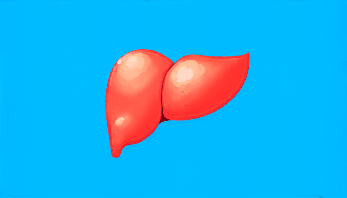
Experiments conducted using the 18F-FDG and 68Ga-FAPI datasets demonstrated that LDOS outperformed state-of-the-art methods in terms of mean Dice scores. The method achieved notable improvements in segmentation accuracy for certain organs, such as the liver and kidneys.
The development of LDOS has significant implications for cancer diagnosis and treatment. Accurate organ segmentation is crucial for quantifying tumor uptake and kinetic measurements, which are essential for monitoring treatment response and predicting patient outcomes.
LDOS also offers a more practical solution for clinical adoption, as it eliminates the need for co-registered CT annotations. This means that clinicians can use LDOS without requiring additional resources or expertise in CT imaging.
While there is still much work to be done in refining LDOS, this new method represents an important step forward in improving the accuracy and efficiency of organ segmentation in PET scans. As researchers continue to develop and refine LDOS, it has the potential to revolutionize cancer diagnosis and treatment, ultimately leading to better patient outcomes and improved quality of life.
Cite this article: “Unlocking Ultra-Low-Dose PET Imaging: A Novel Approach to Organ Segmentation and Quantification”, The Science Archive, 2025.
Positron Emission Tomography, Pet Scans, Organ Segmentation, Low-Dose Pet Images, Denoising, Segmentation, Neural Networks, Masked Autoencoders, Ct-Derived Annotations, Cancer Diagnosis.


