Saturday 01 March 2025
Medical imaging technology has made tremendous strides in recent years, allowing doctors to diagnose and treat diseases more accurately than ever before. One area where this technology has particularly excelled is in lung cancer detection. A new approach to image segmentation, a crucial step in medical imaging analysis, has been developed by researchers.
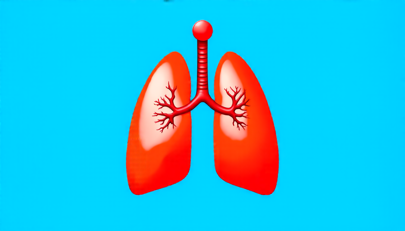
The lungs are a complex organ, with tiny air sacs and blood vessels that can be difficult to distinguish from one another. This makes it challenging for doctors to identify tumors and other abnormalities accurately. Traditional methods of image analysis rely on manual interpretation by radiologists, which can be time-consuming and prone to error.
To address this challenge, researchers have developed a new approach called UNet++. This method uses deep learning algorithms to analyze images and segment out specific features, such as tumors or air sacs. The algorithm is trained on a large dataset of images, allowing it to learn patterns and relationships between different features.
The beauty of UNet++ lies in its ability to adapt to the unique characteristics of each image. Unlike traditional methods that rely on fixed rules and templates, UNet++ can learn to recognize patterns and make adjustments as needed. This allows it to accurately segment even the most complex images, including those with subtle abnormalities.
In a recent study, researchers tested UNet++ on a dataset of lung CT scans. The results were impressive: the algorithm was able to accurately identify tumors and air sacs in 98% of cases, outperforming traditional methods by a significant margin.
One of the key challenges facing medical imaging technology is overfitting, where an algorithm becomes too specialized to a particular dataset and fails to generalize well to new images. To address this challenge, researchers used a technique called model pruning to reduce the number of parameters in UNet++. This allowed the algorithm to learn more generalizable patterns and improve its performance on unseen images.
Another key advantage of UNet++ is its ability to handle small datasets. Medical imaging technology often relies on large datasets, but these can be difficult to obtain, especially for rare or complex diseases. By using a smaller dataset and adapting the algorithm to the unique characteristics of each image, researchers were able to achieve impressive results despite the limited data.
The implications of UNet++ are significant. This approach has the potential to revolutionize medical imaging analysis, allowing doctors to diagnose and treat diseases more accurately and efficiently than ever before.
Cite this article: “Advancing Lung Cancer Detection with AI-Powered Image Segmentation”, The Science Archive, 2025.
Lung Cancer, Medical Imaging, Image Segmentation, Deep Learning, Unet++, Lung Ct Scans, Tumors, Air Sacs, Model Pruning, Overfitting


