
Sunday 02 March 2025
The field of medical image segmentation has seen significant advancements in recent years, driven by the development of deep learning models and their ability to accurately identify various structures within images. However, there is still a need for further improvement, particularly when it comes to small organs and complex anatomical details.
To address this challenge, researchers have turned to radiomics, the high-throughput extraction of quantitative features from medical images. By integrating these features into deep learning models, they hope to create more accurate segmentation algorithms that can better capture subtle changes in image intensity and texture.
One such approach is the GLoG- CSUnet model, which combines a transformer architecture with adaptable Gabor and Laplacian of Gaussian (LoG) filters. These filters are designed to capture multi-scale and multi-orientation texture information, as well as edge detection and boundary precision. By integrating these features into the transformer framework, the GLoG-CSUnet model is able to effectively segment small organs and complex anatomical structures.
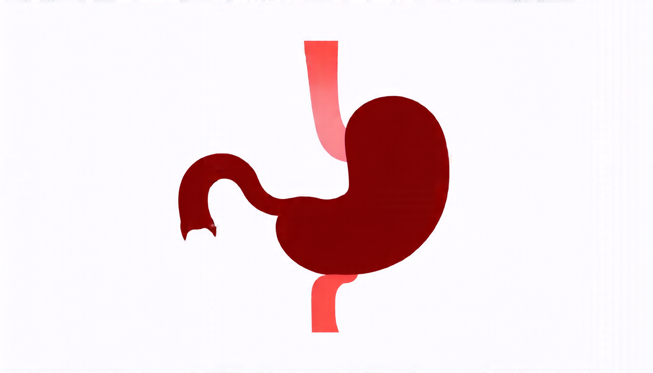
The effectiveness of this approach was tested on two publicly available datasets: the Synapse multi-organ segmentation dataset and the ACDC cardiac segmentation dataset. The results showed that the GLoG-CSUnet model outperformed state-of-the-art methods in both datasets, achieving superior performance in segmenting small organs such as the stomach and pancreas.
One of the key advantages of this approach is its ability to capture local spatial information, which is critical for accurate segmentation of anatomical structures. Traditional convolutional neural networks (CNNs) rely on local receptive fields to extract features, but these can be limited in their ability to capture global context. The transformer architecture used in the GLoG-CSUnet model, on the other hand, is designed to model long-range dependencies between image patches, allowing it to capture more nuanced and complex patterns.
The use of adaptable filters is also a key innovation, as it allows the model to optimize feature extraction for each specific segmentation task. This is particularly important in medical imaging, where different organs and anatomical structures require distinct processing techniques.
While this approach shows significant promise, there are still several challenges that need to be addressed. For example, the authors note that the model’s performance can degrade when applied to small datasets without extensive pre-training. Additionally, the interpretability of the learned filters is an area that requires further exploration.
Cite this article: “Advancing Medical Image Segmentation with Radiomics and Transformers”, The Science Archive, 2025.
Medical Image Segmentation, Deep Learning, Radiomics, Glog-Csunet, Transformer Architecture, Adaptable Filters, Convolutional Neural Networks, Cnns, Medical Imaging, Small Organs, Complex Anatomical Structures