Sunday 02 March 2025
Scientists have long been fascinated by the human body’s intricate networks of fibers and tissues. Now, a team of researchers has developed a revolutionary new imaging technique that allows them to visualize these delicate structures in unprecedented detail.
The method, known as anisotropic X-ray dark-field tomography (AXDT), uses a unique combination of X-rays and computer algorithms to create detailed 3D images of the body’s fibers. Unlike traditional medical imaging techniques, which rely on X-rays or magnetic fields to produce 2D images, AXDT is capable of capturing the intricate details of fibers and tissues in three dimensions.
The technique works by firing a beam of X-rays through the body at a precise angle, creating a diffraction pattern that reveals the orientation and structure of the fibers. This information is then used to reconstruct detailed 3D images of the fibers, allowing researchers to study their shape, size, and arrangement with unprecedented precision.
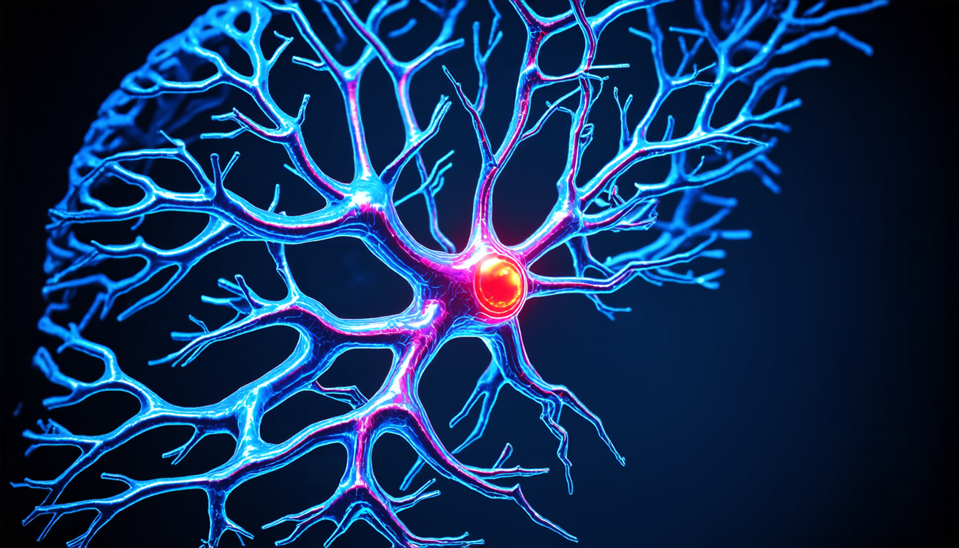
One of the key advantages of AXDT is its ability to visualize fibers in high-resolution, even in areas where traditional imaging techniques struggle to capture detail. For example, the technique has been used to image the delicate fibers in the brain, which are thought to play a crucial role in neurological disorders such as Alzheimer’s disease.
In addition to its potential applications in medicine, AXDT also has exciting implications for materials science and engineering. By allowing researchers to study the internal structure of complex materials, such as fiber-reinforced polymers, the technique could revolutionize our understanding of how these materials behave under stress and strain.
The development of AXDT is a testament to the power of interdisciplinary collaboration, bringing together experts in fields ranging from physics and computer science to medicine and engineering. As researchers continue to refine the technique, it’s likely that we’ll see a wide range of new applications emerge, from improving our understanding of disease mechanisms to developing more efficient and durable materials.
One of the most promising areas of research is in the field of neurological disorders, where AXDT has shown great potential for imaging the delicate fibers in the brain. By allowing researchers to study these fibers in high-resolution, the technique could provide new insights into the underlying causes of diseases such as Alzheimer’s, Parkinson’s, and multiple sclerosis.
In addition to its medical applications, AXDT also has exciting implications for materials science and engineering. For example, the technique could be used to study the internal structure of fiber-reinforced polymers, which are commonly used in aircraft and automotive manufacturing.
Cite this article: “Revolutionary Imaging Technique Reveals Bodys Intricate Fibers in Unprecedented Detail”, The Science Archive, 2025.
X-Ray Imaging, Fibers, Tissues, 3D Imaging, Medical Imaging, Materials Science, Engineering, Computer Algorithms, Tomography, Neuroscience


