Monday 03 March 2025
A new approach has been developed to improve the accuracy of automated retinal vessel segmentation, a crucial technique for diagnosing diseases such as diabetes and glaucoma. The method combines two innovative strategies: structural augmentation and style augmentation.
Retinal vessels are intricate networks that play a vital role in delivering oxygen and nutrients to the retina. However, changes in these vessels can be indicative of underlying health issues. To identify these changes, medical professionals rely on retinal fundus images, which require manual segmentation by experts. This process is time-consuming and prone to human error.
Automated vessel segmentation using deep learning algorithms has shown promising results, but these methods often struggle when applied to new, unseen datasets due to differences in imaging devices and patient demographics. The proposed approach aims to address this issue by enhancing model generalization capabilities.
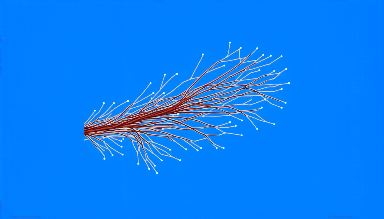
The first component of the method involves structural augmentation, which generates diverse vascular-like structures that mimic actual retinal vessels. These synthetic images are then used to train a Pix2Pix model, allowing the segmentation model to learn a broader range of structure distributions.
The second component is style augmentation, achieved through random photometric augmentations and uncertainty perturbations. This enriches stylistic diversity, enabling the model to adapt better to varying imaging conditions.
The authors evaluated their approach on four challenging datasets: DRIVE, CHASEDB, HRF, and STARE. The results demonstrate state-of-the-art performance, surpassing existing methods. This indicates that the proposed technique can effectively improve the accuracy of automated retinal vessel segmentation.
To generate these synthetic images, the researchers employed a space colonization algorithm, which models the growth of vascular structures in a similar manner to how actual retinal vessels develop. This innovative approach allows for the creation of highly realistic and varied vascular patterns.
The authors also used PixMix to implement random photometric augmentations, introducing uncertainty perturbations that simulate real-world variations in imaging conditions. This adaptation enables the model to learn from diverse datasets and generalize better to unseen data.
In addition to its application in retinal vessel segmentation, this approach has broader implications for medical image analysis. The ability to generate realistic synthetic images can be applied to various fields, such as MRI pulse sequence synthesis or computer-aided detection of lesions.
The development of this method highlights the potential of combining innovative strategies to improve model generalization and accuracy.
Cite this article: “Enhancing Retinal Vessel Segmentation with Structural and Style Augmentations”, The Science Archive, 2025.
Retinal Vessel Segmentation, Automated Segmentation, Deep Learning, Structural Augmentation, Style Augmentation, Pix2Pix Model, Space Colonization Algorithm, Photometric Augmentations, Uncertainty Perturbations, Medical Image Analysis.


