Monday 03 March 2025
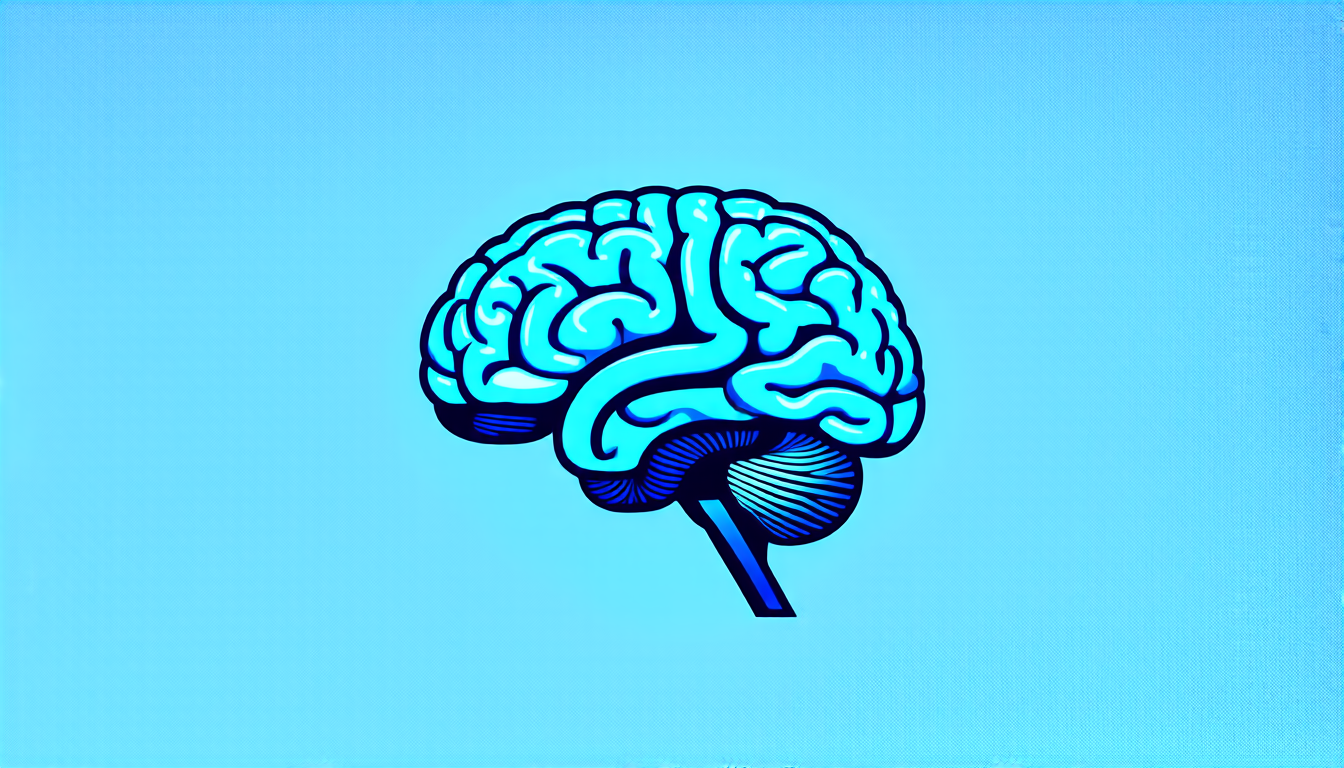
Scientists have long sought a way to peer deeper into the human brain, to better understand its intricate workings and unravel the mysteries of neurological disorders. One major obstacle has been the limited sensitivity of magnetic resonance imaging (MRI) technology, which can struggle to detect subtle changes in brain tissue.
Now, researchers have made a significant breakthrough, developing a new MRI sequence that can produce high-resolution images with unprecedented clarity. This innovation could revolutionize our ability to diagnose and treat neurological conditions such as Alzheimer’s disease, Parkinson’s disease, and stroke.
The new technique, known as True Fast Imaging with Steady-state Precession (True FISP), uses a specialized pulse sequence to amplify the signal from brain tissue. By carefully designing the sequence of radiofrequency pulses, scientists can enhance the signal-to-noise ratio, allowing for more detailed images to be produced.
One key advantage of True FISP is its ability to produce high-resolution images with faster acquisition times than traditional MRI sequences. This is particularly important in neurology, where rapid diagnosis and treatment are crucial. With True FISP, clinicians may be able to quickly identify subtle changes in brain tissue, enabling them to intervene earlier and more effectively.
The new sequence has already been tested on a range of subjects, including healthy volunteers and patients with neurological disorders. The results have been striking, with high-resolution images that reveal previously unseen details about the brain’s structure and function.
For example, researchers were able to produce detailed images of the brain’s white matter tracts, which are critical for communication between different regions of the brain. These images could help clinicians better understand how neurological disorders affect these tracts, and develop more targeted treatments.
Another area where True FISP has shown promise is in the study of brain development and aging. By producing high-resolution images of the brain’s structure and function over time, researchers may be able to identify early signs of neurodegenerative disease or cognitive decline.
The potential implications of this technology are vast. With True FISP, clinicians could potentially use MRI to diagnose neurological disorders at an earlier stage, when treatment is more effective. They could also use the technique to monitor the progression of disease and track the response to treatment.
Of course, there are still many challenges to overcome before True FISP becomes a clinical reality. The technology will need to be refined and optimized for use in a variety of settings, and clinicians will need to be trained on how to interpret the resulting images.
Still, the potential benefits of this innovation are clear.
Cite this article: “Breakthrough MRI Sequence Revolutionizes Neurological Imaging”, The Science Archive, 2025.
Magnetic Resonance Imaging, Neurology, Alzheimer’S Disease, Parkinson’S Disease, Stroke, True Fisp, Mri Sequence, Brain Tissue, High-Resolution Images, Neurological Disorders