
Tuesday 04 March 2025
Cells are the building blocks of life, and understanding how they move and interact is crucial for unlocking new insights into biological processes and developing novel treatments for diseases. In a fascinating study, researchers have shed light on the dynamics of cell migration in epithelial cell monolayers, revealing that defects in cell shape can drive collective motion.
Epithelial cells form a layer of cells that lines the inner surfaces of organs, glands, and other tissues. When these cells move collectively, they play crucial roles in embryonic development, tissue repair, and disease progression. However, the underlying mechanisms that govern this movement remain poorly understood.
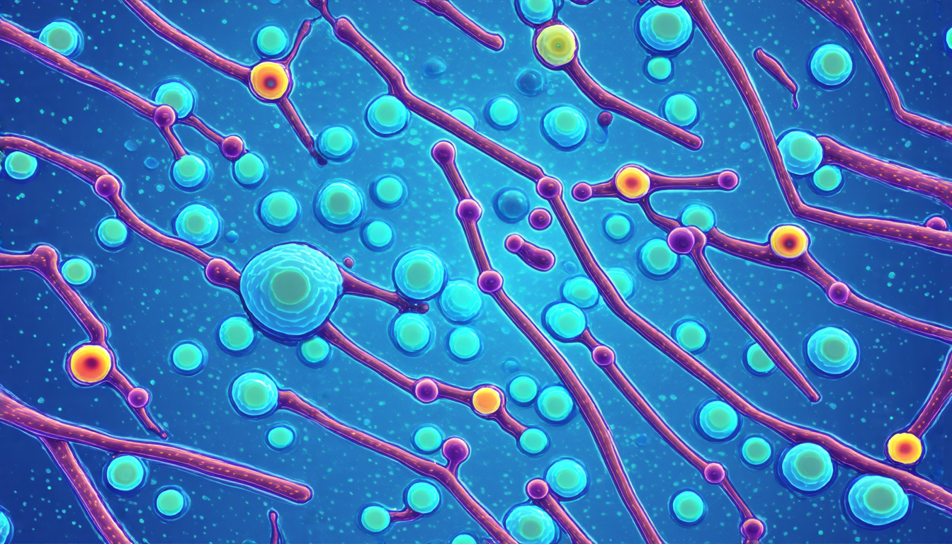
The researchers studied epithelial cell monolayers from Madin-Darby canine kidney (MDCK) cells, which are commonly used as a model system for understanding cellular behavior. They observed that when these cells move collectively, defects in cell shape can emerge, leading to the formation of topological defects. These defects can take two forms: head-to-tail and tail-to-head.
The researchers used advanced imaging techniques to visualize the movement of cells and defects over time. They found that both types of defects exhibit distinct patterns of motion, with head-to-tail defects moving in a continuous, smooth manner, while tail-to-head defects move in a more erratic fashion.
Further analysis revealed that the defects are driven by the interaction between cell forces and stresses within the epithelial layer. The researchers discovered that the cells exert traction forces on their substrate, which are balanced by stresses within the layer. These stresses can either amplify or dampen the motion of the defects, depending on the orientation of the cells.
The study also explored the role of stress fibers in regulating cell migration. Stress fibers are protein networks that provide mechanical support to cells and play a crucial role in cellular movement. The researchers found that stress fibers near topological defects exhibit specific orientations, which are influenced by the motion of the defects.
These findings have important implications for our understanding of epithelial cell migration and its role in biological processes. The study highlights the complex interplay between cell forces, stresses, and shape defects in driving collective motion. Future research can build upon these discoveries to uncover new insights into cellular behavior and develop novel therapeutic strategies for diseases related to defective cell migration.
The researchers’ work demonstrates the power of interdisciplinary approaches that combine advanced imaging techniques with mathematical modeling to understand biological phenomena.
Cite this article: “Unraveling the Dynamics of Epithelial Cell Migration: A Study on Collective Motion and Defects”, The Science Archive, 2025.
Cell Migration, Epithelial Cells, Collective Motion, Cell Shape, Topological Defects, Stress Fibers, Traction Forces, Stresses, Biological Processes, Mathematical Modeling