
Wednesday 05 March 2025
Ultrasound technology has come a long way since its inception in the early 20th century. From its humble beginnings as a tool for medical diagnosis, it has evolved into a powerful means of visualizing internal organs and tissues. In recent years, researchers have made significant strides in improving the resolution and quality of ultrasound images.
One such advancement is the development of a new beamforming algorithm called SAMAS (Sub-Aperture Angular Multiply and Sum). This innovative technique combines the advantages of two existing methods – FMAS (Frame-Multiply-and-Sum) and ASAP (Acoustic Sub-Aperture Pairing) – to produce high-resolution images with reduced noise.
The key challenge in ultrasound imaging is separating the weak signals from moving blood vessels from the strong echoes from surrounding tissues. Traditional beamforming techniques, such as Delay-and-Sum (DAS), struggle to achieve this distinction, resulting in low-quality images with limited detail.
SAMAS addresses these limitations by incorporating two main components: frame-multiply-and-sum and acoustic sub-aperture pairing. The former involves multiplying the signals from multiple angles and summing them to enhance the contrast between blood vessels and surrounding tissues. The latter involves splitting the data into smaller sub-arrays, which are then processed separately to reduce noise.
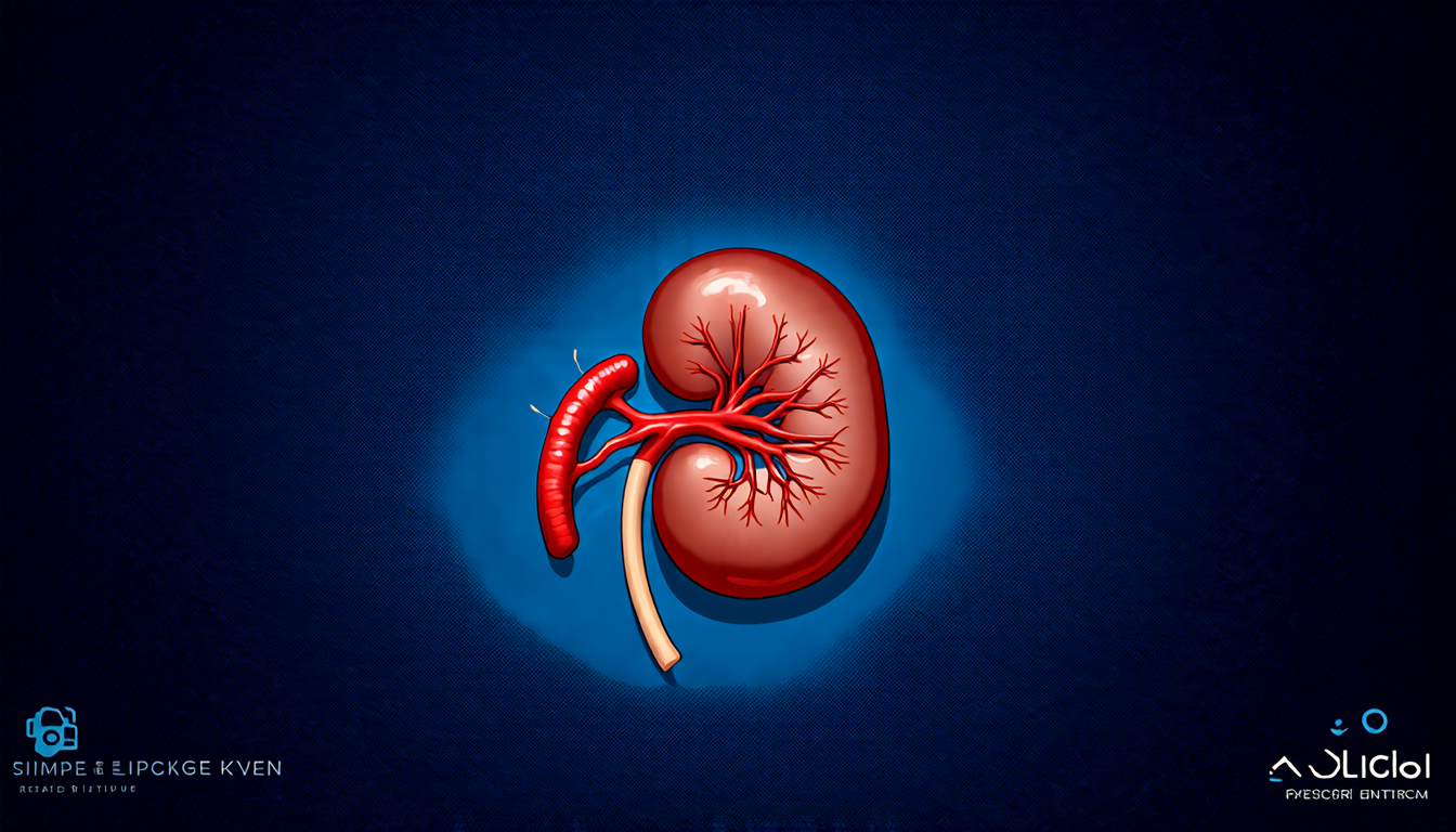
In a recent study, researchers tested SAMAS on both in vitro and in vivo experiments using a rabbit kidney model and human lymph node imaging. The results were astounding – SAMAS consistently improved the contrast-to-noise ratio (CNR) and signal-to-noise ratio (SNR) compared to traditional beamforming techniques.
One of the most significant benefits of SAMAS is its ability to enhance the visibility of small blood vessels, which are crucial for diagnosing certain medical conditions. By reducing noise and increasing resolution, SAMAS allows clinicians to visualize these tiny vessels with unprecedented clarity.
Another advantage of SAMAS is its flexibility – it can be applied to a wide range of ultrasound imaging applications, from cardiovascular disease to cancer diagnosis. This versatility makes it an attractive option for researchers and clinicians alike.
While SAMAS shows tremendous promise, there are still some limitations to consider. For example, the algorithm requires significant computational power, which may pose challenges for real-time processing. Additionally, further testing is needed to assess its effectiveness in different clinical scenarios.
Despite these challenges, the development of SAMAS represents a major breakthrough in ultrasound imaging technology.
Cite this article: “Advancing Ultrasound Imaging with SAMAS Technology”, The Science Archive, 2025.
Ultrasound, Imaging, Beamforming, Samas, Algorithm, Resolution, Quality, Noise, Blood Vessels, Medical Diagnosis.