

Monday 10 March 2025
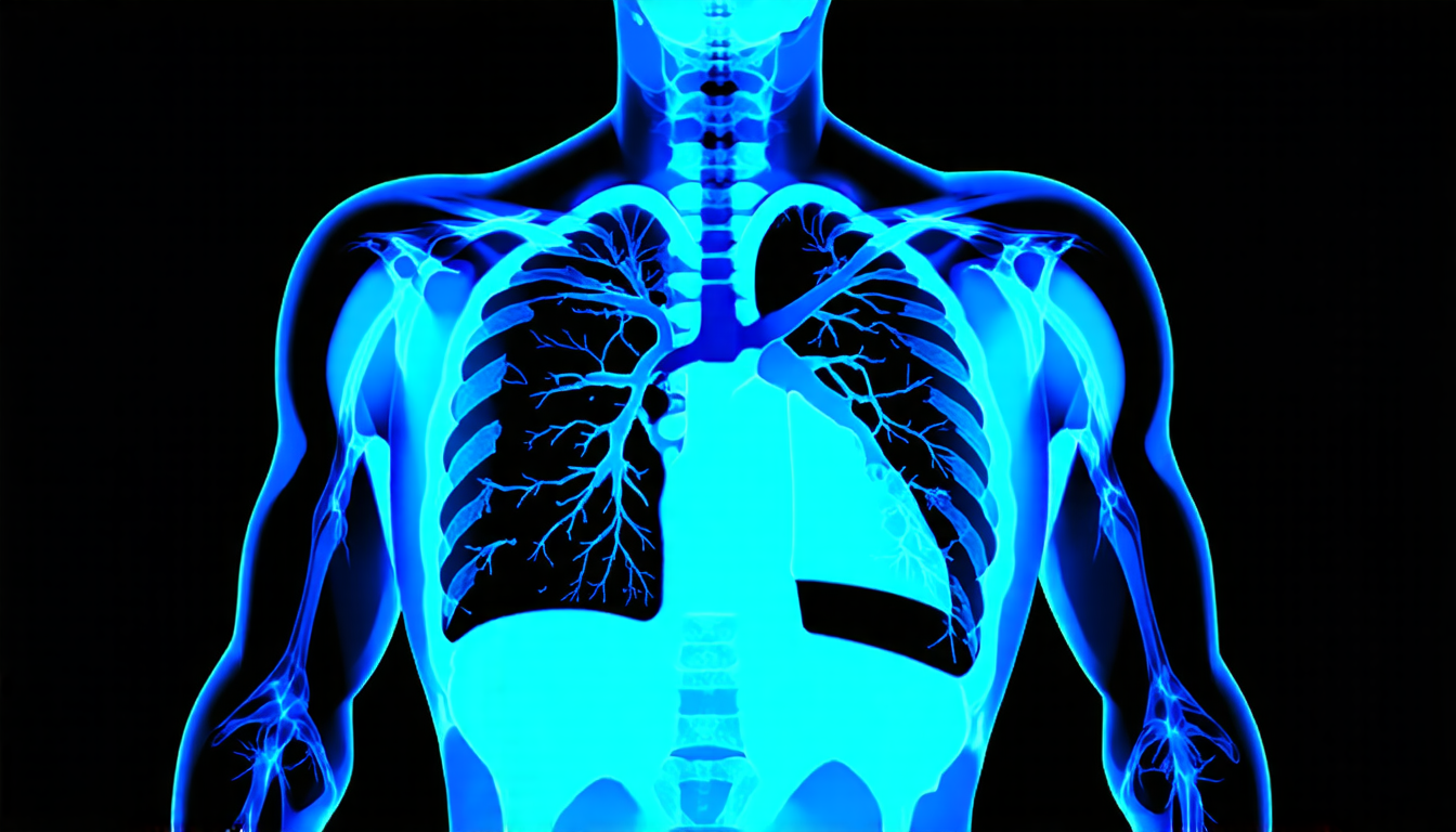
A team of researchers has made significant strides in developing a new approach for medical image analysis, which could revolutionize the way doctors diagnose and treat patients. By leveraging large language models and fine-grained annotations, the method is capable of accurately recognizing subtle patterns and details in chest X-ray images that are often overlooked by traditional computer vision techniques.
The researchers’ approach, known as MedFILIP, uses a technique called contrastive learning to train a model on paired images and text data. This allows the model to learn rich representations of medical concepts and visual attributes, enabling it to identify subtle patterns and relationships between them.
One of the key innovations behind MedFILIP is its ability to extract fine-grained entities from diagnosis reports, such as disease categories, severity levels, and locations. By decoupling this information from the text, the model can learn to recognize specific visual attributes associated with each entity, allowing it to make more accurate diagnoses.
To test the effectiveness of MedFILIP, the researchers used a range of datasets, including publicly available chest X-ray images and reports from various hospitals. They found that their method outperformed traditional computer vision techniques in recognizing subtle patterns and details, such as pneumonia and other lung diseases.
The potential applications of MedFILIP are vast. By enabling doctors to quickly and accurately diagnose patients with conditions like pneumonia, the method could help reduce wait times and improve patient outcomes. Additionally, MedFILIP’s ability to learn from large datasets of paired images and text data makes it an attractive tool for researchers seeking to develop new medical imaging techniques.
The development of MedFILIP is a testament to the power of collaboration between computer scientists, medical professionals, and engineers. By combining their expertise, the researchers have created a cutting-edge approach that has the potential to transform the field of medical image analysis.
In the future, the team plans to continue refining MedFILIP and exploring its applications in other areas of medicine. As the method continues to evolve, it could ultimately lead to more accurate diagnoses, improved patient care, and new breakthroughs in medical research.
Cite this article: “Revolutionizing Medical Image Analysis with MedFILIP”, The Science Archive, 2025.
Medical Image Analysis, Deep Learning, Chest X-Ray Images, Diagnosis Reports, Contrastive Learning, Fine-Grained Entities, Lung Diseases, Pneumonia, Medical Imaging Techniques, Natural Language Processing





