Thursday 23 January 2025
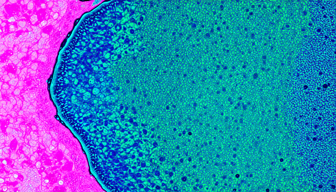
The quest for a more efficient way to analyze whole slide images (WSIs) in digital pathology has led scientists to develop a novel approach that combines patch-based feature extraction, K-means clustering, and Fisher vector encoding. This method, presented in a recent study, shows great promise in classifying WSIs with high accuracy.
In traditional machine learning models, the sheer size and complexity of WSIs can pose significant computational challenges. One common solution is to divide the images into smaller patches and process them independently. However, this approach may overlook broader structural information, which is crucial for accurate diagnosis.
The new method tackles this issue by extracting features from each patch using pre-trained convolutional neural networks (CNNs) or transformers. These features are then clustered using K-means clustering, reducing the dimensionality of the feature space while preserving important tissue heterogeneity.
To further compress the data, the study uses Fisher vector encoding to model the distribution of feature embeddings within each cluster. This results in a compact representation of the entire WSI that captures both local and global information.
The researchers tested their method on various datasets, including HER2 scoring, HER2+ vs HER2- classification, EGFR mutation prediction, and metastasis detection. The results show that the proposed approach outperformed traditional patch-based methods across all tasks, demonstrating its robustness and scalability.
One of the key advantages of this method is its ability to adapt to different imaging modalities and classification tasks. This versatility is particularly valuable in digital pathology, where diverse datasets and applications are common.
The study’s findings also highlight the potential for deep learning models to improve diagnostic accuracy in digital pathology. By combining patch-based feature extraction with clustering and Fisher vector encoding, researchers can create a more comprehensive representation of WSIs that captures both local and global features.
While the results are promising, further work is needed to assess the method’s performance in real-world clinical settings. Nevertheless, this innovative approach has the potential to revolutionize the field of digital pathology, enabling faster and more accurate diagnosis of diseases.
In addition to its technical merits, the study showcases the power of interdisciplinary collaboration between computer scientists, biologists, and clinicians. By combining expertise from multiple fields, researchers can develop novel solutions that address complex challenges in healthcare.
Cite this article: “Novel Approach to Analyzing Whole Slide Images in Digital Pathology”, The Science Archive, 2025.
Whole Slide Images, Digital Pathology, Machine Learning, Patch-Based Feature Extraction, K-Means Clustering, Fisher Vector Encoding, Convolutional Neural Networks, Transformers, Image Classification, Deep Learning.


