

Wednesday 12 March 2025
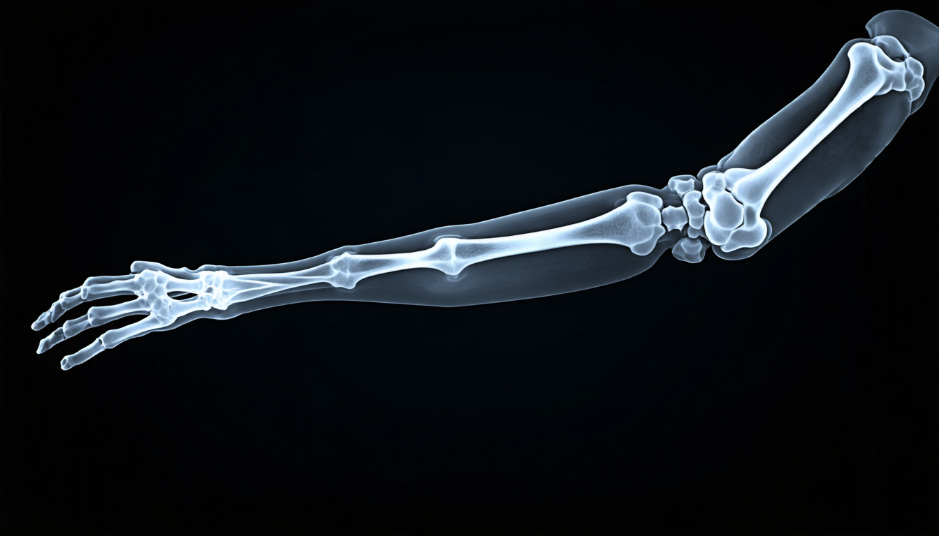
X-ray images are a vital tool in medical diagnosis, helping doctors identify and treat a wide range of conditions. However, these images can be notoriously tricky to interpret, particularly when they’re not presented clearly. For radiologists, the challenge is compounded by the fact that different patients have different anatomies, scanning positions, and patient sizes – all of which can affect how an image looks.
In recent years, deep learning has revolutionized medical imaging analysis, allowing researchers to develop sophisticated algorithms that can automatically correct for these variations and enhance image quality. But while these methods are undeniably powerful, they often rely on complex, opaque models that can be difficult for clinicians to understand or interpret.
Now, a team of researchers from GE Healthcare has proposed a new approach to X-ray image enhancement that aims to change this status quo. By decomposing the traditional image processing workflow into a series of interpretable, pixel-level operations, their method provides radiologists with a clear understanding of how images are being enhanced – and why.
The key innovation lies in the team’s use of a deep learning model inspired by the multi-level image processing workflow used in conventional X-ray enhancement algorithms. This approach allows the researchers to predict not just global brightness and contrast adjustments, but also regional LUTs (lookup tables) that can adapt to specific anatomies and scanning positions.
In other words, their method is designed to mimic the way radiologists would manually adjust X-ray images – but does so automatically, using data-driven insights to guide its decisions. This not only improves image quality, but also provides clinicians with a level of transparency and control they’ve never had before.
To test their approach, the researchers trained their model on a dataset of 429 clinical X-ray images from around the world, each annotated with correct parameters for brightness and contrast adjustment. They then compared the performance of their method to several state-of-the-art algorithms, including ResUNet, DLBC (deep learning-based brightness and contrast correction), 3DLUT, and Sup-DCE.
The results are impressive: not only does their method achieve higher PSNR (peak signal-to-noise ratio) scores than its competitors, but it also produces images with more consistent presentations across different anatomies and scanning positions. In other words, the enhanced X-ray images look more like they were taken from a single, idealized patient – rather than a diverse group of individuals.
Cite this article: “Enhancing Transparency and Control in X-ray Image Analysis”, The Science Archive, 2025.
X-Ray Images, Medical Diagnosis, Deep Learning, Image Enhancement, Radiologists, Anatomies, Scanning Positions, Patient Sizes, Interpretable Models, Opaque Models.





