

Thursday 13 March 2025
The development of artificial intelligence (AI) has led to numerous breakthroughs in various fields, including medicine. In recent years, researchers have been working on creating AI models that can accurately diagnose and classify diseases using medical images, such as computed tomography (CT) scans and magnetic resonance imaging (MRI).
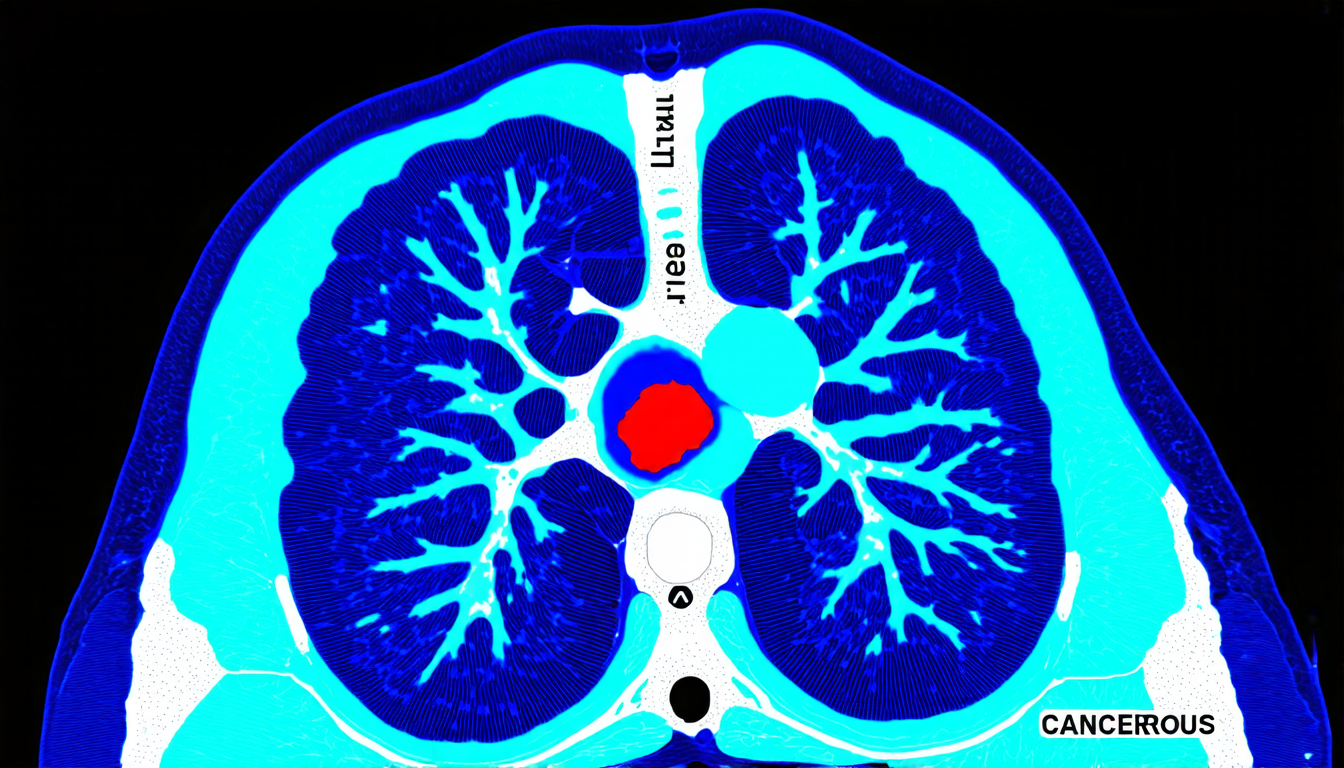
A new study published in a prestigious scientific journal has made significant strides in this area by introducing a novel approach to integrating CT images with clinical data for cancer diagnosis. The researchers used a combination of multiple instance learning (MIL) and contrastive learning to develop a multimodal model that can accurately classify cancer types based on both imaging and clinical features.
The study’s authors began by collecting a large dataset of CT scans and corresponding clinical data from patients with various types of cancer, including breast, lung, and colon cancer. They then used MIL to process the CT images and extract relevant features, such as tumor size and shape. The clinical data was also processed using a similar approach.
The researchers then combined the imaging and clinical features using contrastive learning, which involves training a model to distinguish between positive pairs (i.e., images and clinical data from patients with cancer) and negative pairs (i.e., images and clinical data from patients without cancer). This approach allowed the model to learn a shared representation space for both imaging and clinical data.
The resulting multimodal model was evaluated using multiple classification tasks, including breast cancer staging, lung cancer histology, and colon cancer diagnosis. The results showed that the model outperformed traditional unimodal approaches, which use only imaging or clinical data, in all three tasks.
One of the key advantages of this approach is its ability to leverage the strengths of both imaging and clinical data. Imaging modalities like CT scans provide detailed information about tumor morphology, while clinical data offers insights into patient-specific characteristics, such as age, sex, and medical history. By combining these two types of data, the model can learn more accurate and robust representations of cancer.
The study’s findings have significant implications for the development of AI-powered diagnostic tools in medicine. By integrating multimodal data, researchers can create more accurate and effective models that can improve patient outcomes and reduce the risk of misdiagnosis.
However, there are also challenges to be addressed before this technology can be widely adopted. For example, the availability and quality of medical images and clinical data can vary significantly depending on the location and healthcare system.
Cite this article: “Multimodal AI Model Accurately Diagnoses Cancer Using CT Images and Clinical Data”, The Science Archive, 2025.
Artificial Intelligence, Medical Imaging, Cancer Diagnosis, Multimodal Learning, Contrastive Learning, Computed Tomography Scans, Magnetic Resonance Imaging, Clinical Data, Breast Cancer, Colon Cancer





