

Thursday 13 March 2025
Medical image analysis has long been plagued by a lack of labeled data, making it difficult for AI models to learn and improve. But now, researchers have found a way to overcome this hurdle using clever data augmentation techniques.
The problem is that medical imaging datasets are often small and imbalanced, with a limited number of examples from certain classes or conditions. This can lead to AI models struggling to generalize and make accurate predictions. To combat this, researchers have turned to data augmentation – the process of artificially increasing the size and variability of a dataset by applying transformations to the images.
In a new study, scientists used two different approaches to augment their medical image datasets. The first was traditional data augmentation, which involves applying geometric and photometric transformations to the images, such as rotation, scaling, and brightness adjustment. This is a common technique in computer vision, but it can be limited in its ability to capture complex patterns and relationships.
The second approach was more innovative – the researchers used a technique called Mixup, which combines two images and their corresponding masks to generate new synthetic samples. This allows for the creation of more diverse and realistic training data, which can help AI models learn better features and improve their performance.
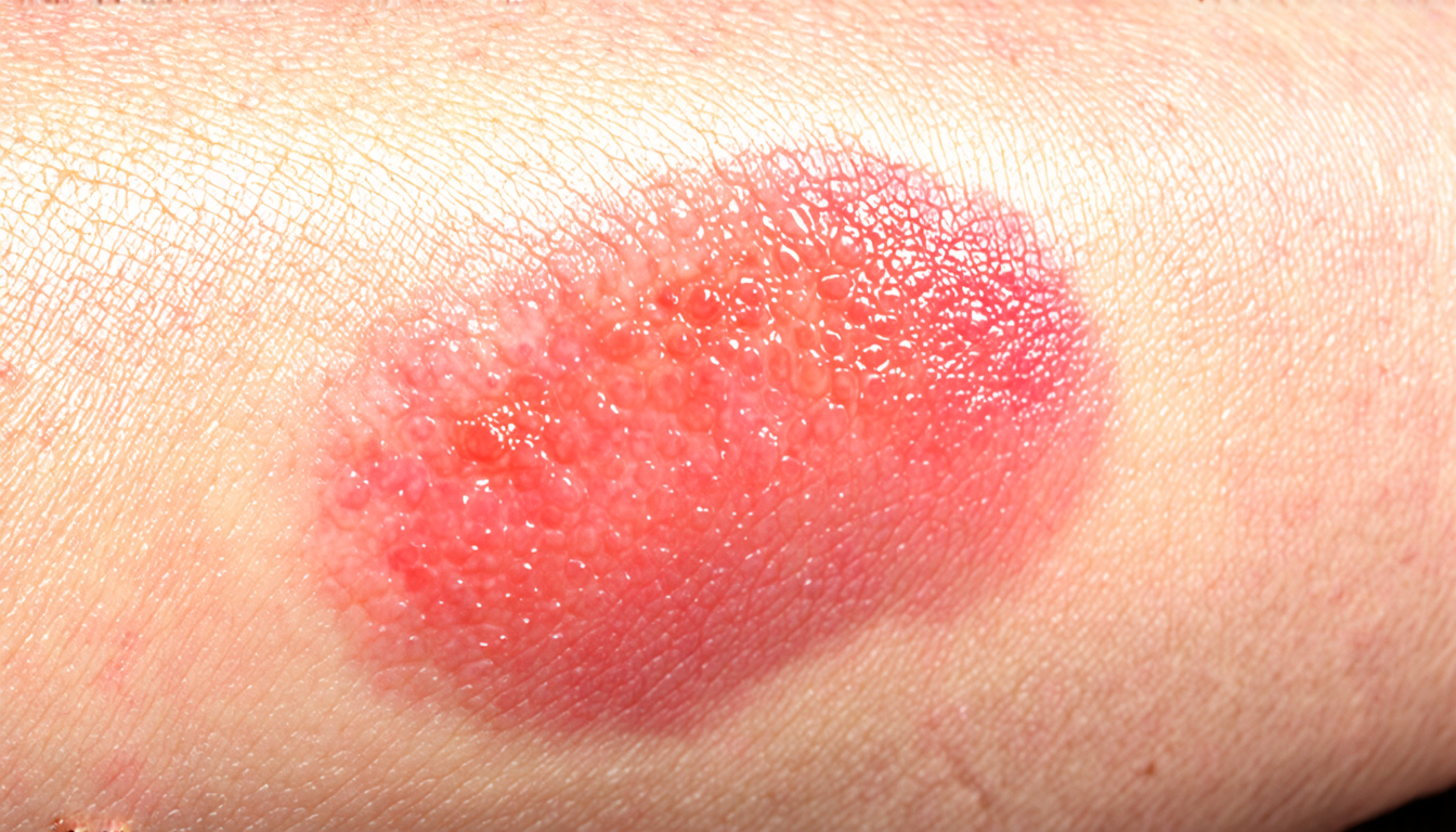
To test these approaches, the researchers used them on two different medical image datasets – one containing skin cancer images, and another showing blood vessels in retinal fundus images. They then trained convolutional neural networks (CNNs) to classify the skin cancer images and segment the blood vessels.
The results were impressive. In the skin cancer classification task, the CNN trained with traditional data augmentation achieved an accuracy of 90.74%, while the model trained with Mixup achieved an accuracy of 96.88%. This represents a significant improvement in performance, demonstrating that Mixup can be effective in improving the robustness and accuracy of AI models.
In the blood vessel segmentation task, the researchers found that traditional data augmentation was unable to improve the model’s performance, while Mixup resulted in a notable improvement in Dice coefficient – a measure of segmentation quality. This suggests that Mixup can help AI models learn more accurate features and make better predictions in medical image analysis tasks.
The findings have important implications for medical imaging research. By using data augmentation techniques like Mixup, researchers may be able to overcome the limitations of small and imbalanced datasets, and develop more accurate and reliable AI models for diagnosis and treatment planning. This could ultimately lead to improved patient outcomes and better healthcare decision-making.
Cite this article: “Overcoming Data Limitations in Medical Image Analysis with Innovative Augmentation Techniques”, The Science Archive, 2025.
Medical Image Analysis, Data Augmentation, Mixup, Convolutional Neural Networks, Cnns, Skin Cancer, Blood Vessels, Retinal Fundus Images, Segmentation, Classification





