Thursday 20 March 2025
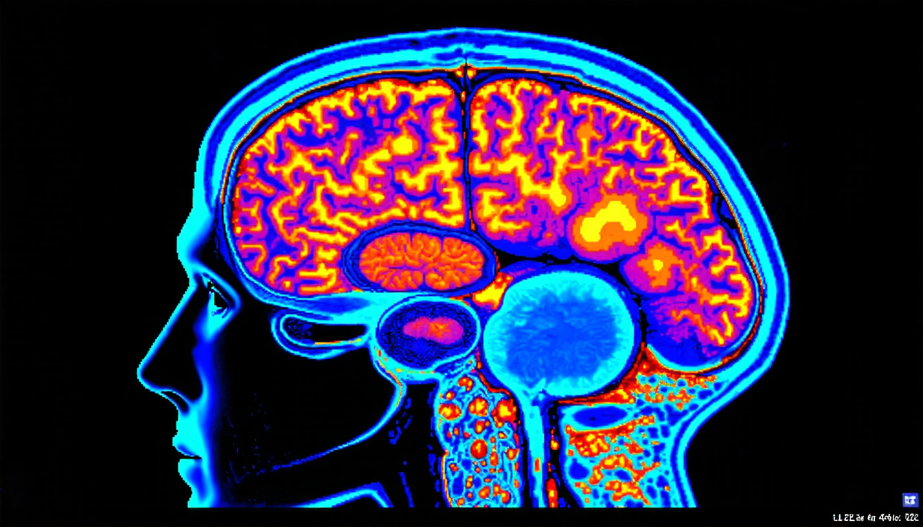
Medical imaging has come a long way in recent years, allowing doctors to see inside the human body like never before. But despite these advances, there’s still one major limitation: the need for contrast agents. These substances are injected into patients’ veins to highlight specific areas of the body, helping doctors diagnose diseases and track their progress.
However, these agents can have serious side effects, such as kidney damage or allergic reactions. So, researchers have been working on a new way to create high-quality medical images without using them. Their solution involves machine learning algorithms and something called dynamic contrast-enhanced magnetic resonance imaging (DCE-MRI).
In DCE-MRI, a strong magnetic field and radio waves are used to take detailed pictures of the body’s internal structures. But unlike traditional MRI scans, which only show the structure of the body, DCE-MRI also measures how blood flows through different areas. This information is crucial for diagnosing conditions like cancer, where tumors often have abnormal blood flow patterns.
The problem is that DCE-MRI requires two separate scans: one without contrast agents and another with them. The second scan can take up to an hour, which isn’t ideal for patients who are already anxious or in pain. So, researchers set out to develop a machine learning algorithm that could synthesize the second scan from the first.
Their approach uses something called a generative adversarial network (GAN), which is essentially two neural networks competing against each other. One network generates images based on the first scan, while the other network tries to distinguish between these generated images and real ones taken with contrast agents.
By training this GAN on a large dataset of MRI scans, researchers were able to create high-quality images that mimic those taken with contrast agents. These images show not only the structure of the body but also the blood flow patterns, making it easier for doctors to diagnose diseases.
But how did they get these results? The key was in incorporating additional information from other types of medical imaging modalities, such as T2-weighted and ADC (apparent diffusion coefficient) maps. These maps provide complementary information about the body’s internal structures, which is essential for creating accurate images.
The researchers also experimented with different attention mechanisms to focus on specific regions of interest, like the prostate gland or brain tumors. By doing so, they were able to create more detailed and accurate images that highlight these areas.
The potential impact of this technology is huge.
Cite this article: “Machine Learning-Powered MRI Technology Revolutionizes Diagnostic Imaging Without Contrast Agents”, The Science Archive, 2025.
Medical Imaging, Machine Learning, Mri Scans, Contrast Agents, Dynamic Contrast-Enhanced Magnetic Resonance Imaging, Generative Adversarial Network, Neural Networks, Cancer Diagnosis, Blood Flow Patterns, Disease Diagnosis.


