

Wednesday 26 March 2025
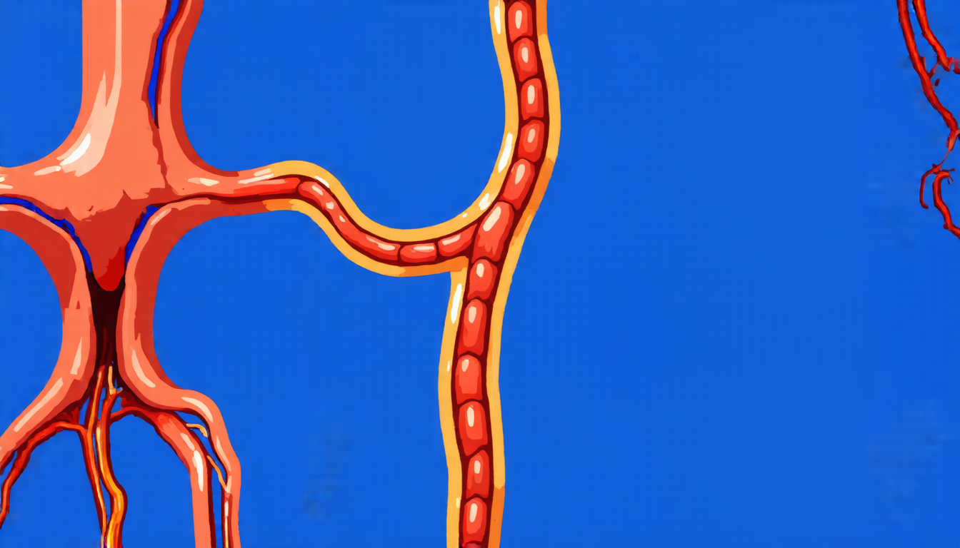
A team of researchers has developed a new approach for segmenting 3D images of the carotid artery, a crucial step in diagnosing and monitoring cardiovascular disease.
The traditional method of segmenting these images involves manually tracing the vessel walls, which is both time-consuming and prone to human error. To overcome this challenge, scientists have turned to machine learning algorithms that can learn patterns from training data and apply them to new images.
However, creating high-quality training data for 3D carotid artery segmentation has been a significant hurdle. Researchers have relied on sparse annotations, which are limited in number and often lack consistency. This limitation has led to suboptimal performance of machine learning algorithms, making accurate diagnosis and monitoring more difficult.
To address this issue, the team developed an iterative approach that uses adversarial networks to create pseudo-labels from sparse annotations. These pseudo-labels are then used to train a 3D convolutional neural network (CNN) for segmentation.
The key innovation is the use of centerline sampling to create densely distributed contours of the carotid artery wall. This allows the CNN to learn patterns and features that were previously hidden in the sparse annotations.
To evaluate the performance of their approach, the researchers used a dataset of 202 MRI volumes from patients with hypertension, cardiovascular risk factors, or plaque in the internal carotid artery. They compared their method to other state-of-the-art approaches and found significant improvements in segmentation accuracy.
The results have important implications for clinical practice. Accurate segmentation of the carotid artery wall can help diagnose atherosclerosis, assess stenosis, and monitor treatment outcomes. By leveraging machine learning algorithms with pseudo-labels created from sparse annotations, researchers hope to improve the efficiency and effectiveness of cardiovascular disease diagnosis and management.
In addition to its potential applications in clinical settings, this work highlights the importance of developing innovative approaches to image segmentation. As medical imaging technologies continue to evolve, it is crucial that researchers develop methods that can keep pace with these advancements.
The team’s approach has also sparked new avenues for research in the field of machine learning and computer vision. By exploring different sampling strategies and network architectures, scientists may uncover even more effective ways to segment 3D images of the carotid artery.
Ultimately, this work demonstrates the power of collaboration between researchers from different disciplines. By combining expertise in medicine, imaging, and machine learning, scientists can develop innovative solutions that transform our understanding of complex diseases like cardiovascular disease.
Cite this article: “Segmenting the Carotid Artery: A New Approach to Accurate Diagnosis and Monitoring”, The Science Archive, 2025.
Machine Learning, 3D Image Segmentation, Carotid Artery, Cardiovascular Disease, Mri Volumes, Hypertension, Atherosclerosis, Stenosis, Computer Vision, Adversarial Networks





