Thursday 27 March 2025
The quest for a more efficient and accurate way of documenting corpses in forensic medicine has led researchers to develop an innovative robotic system that can autonomously scan the surface of a body, providing detailed information about any injuries or wounds.
For decades, forensic pathologists have relied on traditional methods such as photography and manual measurements to document external injuries. However, these approaches are time-consuming, prone to human error, and often lack the level of detail required in modern forensic investigations. To address this issue, a team of researchers has designed a mobile robot that can quickly and accurately capture 3D surface data of a body.
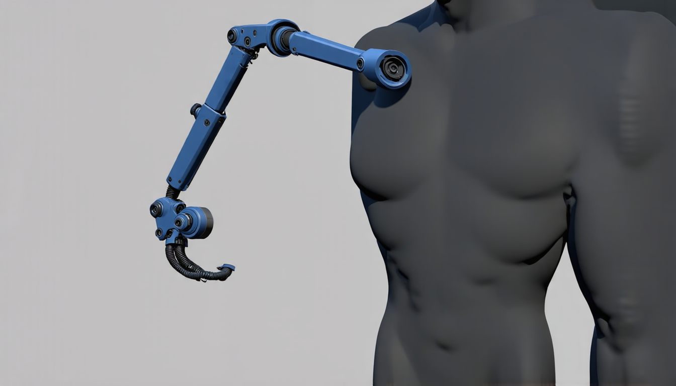
The robotic system consists of a lightweight robotic arm mounted on a differential drive base, which allows it to move freely around the body. An RGB-D camera is attached to the end of the arm, capturing high-resolution images and depth information simultaneously. This combination enables the robot to create a detailed 3D point cloud of the body’s surface.
To optimize its scanning path, the robot uses a configuration space analysis that takes into account various environmental factors such as body geometry, couch height, and workspace constraints. The system can adapt to different settings by adjusting its scanning route accordingly.
In a series of experiments, the robotic system was tested in a laboratory setting using a male body phantom and in a forensic CT room with real corpses. Results showed that the robot could achieve high levels of surface coverage, even in confined spaces, and accurately capture detailed information about injuries and wounds.
One of the key benefits of this robotic approach is its ability to reduce the time and effort required for documentation, allowing pathologists to focus on more complex aspects of their work. Additionally, the system’s autonomous nature minimizes the risk of human error and contamination, ensuring that evidence is preserved and handled properly.
The potential applications of this technology extend beyond forensic medicine. In medical education, it could be used to create realistic simulation models for training purposes. In clinical practice, it could aid in wound assessment and treatment planning.
While there are still some limitations to the system, such as the need for manual post-processing to stitch together the captured point clouds, these challenges can be addressed through further development and refinement.
As forensic medicine continues to evolve, innovations like this robotic scanning system will play a crucial role in improving the accuracy and efficiency of investigations. By providing more detailed and reliable information about injuries and wounds, it has the potential to make a significant impact on the field and ultimately help bring justice to victims and their families.
Cite this article: “Autonomous Robotic System for Accurate Forensic Documentation of Corpses”, The Science Archive, 2025.
Forensic Medicine, Robotics, Scanning System, 3D Point Cloud, Rgb-D Camera, Configuration Space Analysis, Surface Data, Injury Documentation, Wound Assessment, Medical Education


