Friday 28 March 2025
Deep learning has revolutionized many areas of science and technology, but its applications in medical imaging have been particularly impressive. One of the most significant challenges in medical imaging is reconstructing images using different kernels, which can be thought of as filters that enhance or suppress certain features in the image.
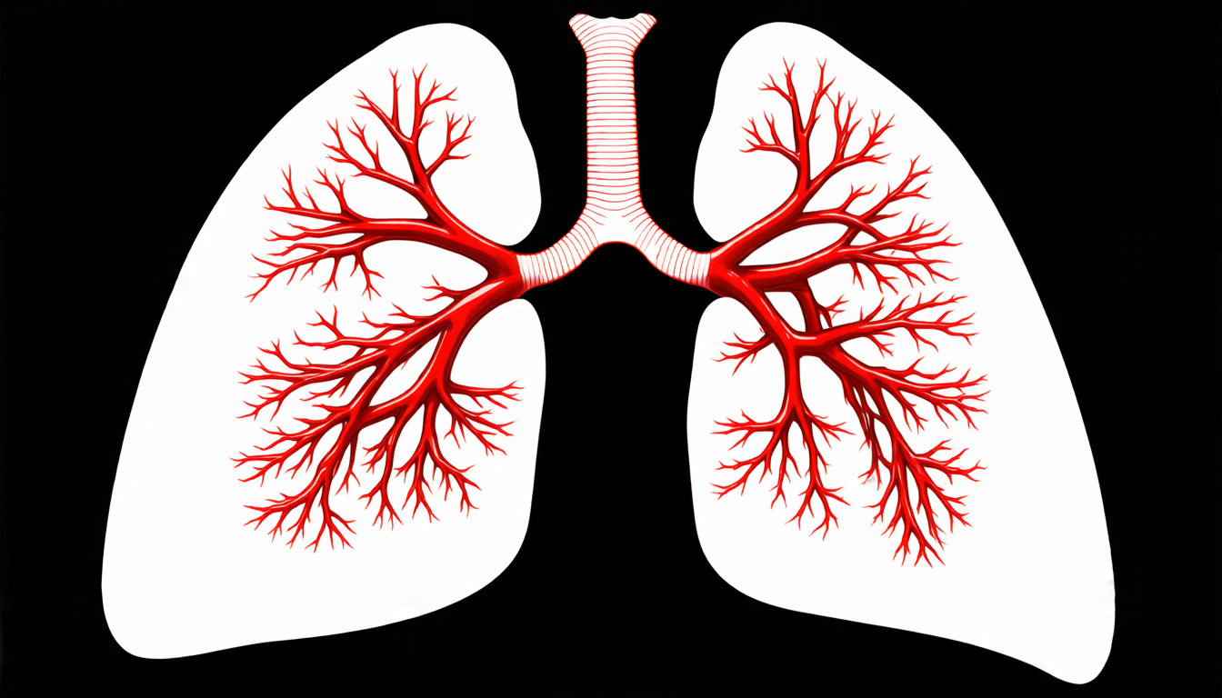
Reconstructing images using different kernels is crucial in medical imaging because it allows doctors to see the same patient’s anatomy from different angles and with varying levels of detail. For example, a smooth kernel can be used to highlight soft tissue structures like lungs and liver, while a sharp kernel can be used to emphasize bone and vessel details.
However, reconstructing images using different kernels is not as simple as just switching between two different filters. The problem is that the data acquired by the CT scanner is not perfect, and it’s affected by factors like noise and artifacts. This means that simply applying a different kernel to the raw data can produce poor-quality images with distorted features.
To overcome this challenge, researchers have developed a new deep learning-based approach that uses model-based deep learning to reconstruct images using different kernels. The key innovation is the use of a forward model that incorporates information about the kernel’s modulation transfer function (MTF), which describes how the kernel affects the image.
The MTF is a critical component in this approach because it allows the network to learn how to correct for the effects of noise and artifacts on the raw data. By incorporating the MTF into the forward model, the network can produce images that are more accurate and detailed than those produced by traditional methods.
One of the most impressive features of this new approach is its ability to reconstruct images using different kernels with varying levels of detail. For example, a kernel with a higher frequency cut-off can be used to highlight fine details like blood vessels, while a kernel with a lower frequency cut-off can be used to emphasize larger structures like bones.
The researchers tested their approach using clinical data from the GE Revolution Ascend system and found that it produced high-quality images with improved detail and contrast. They also compared their results to those produced by a direct learning method, which did not incorporate information about the kernel’s MTF. The results showed that the new approach was able to produce sharper images with less noise and artifacts.
This new approach has significant implications for medical imaging, particularly in the diagnosis and treatment of diseases like cancer.
Cite this article: “Deep Learning-Based Image Reconstruction for Medical Imaging Applications”, The Science Archive, 2025.
Medical Imaging, Deep Learning, Kernel Filters, Image Reconstruction, Ct Scanner, Noise Reduction, Artifact Correction, Modulation Transfer Function (Mtf), Model-Based Deep Learning, Image Quality Improvement


