Friday 28 March 2025
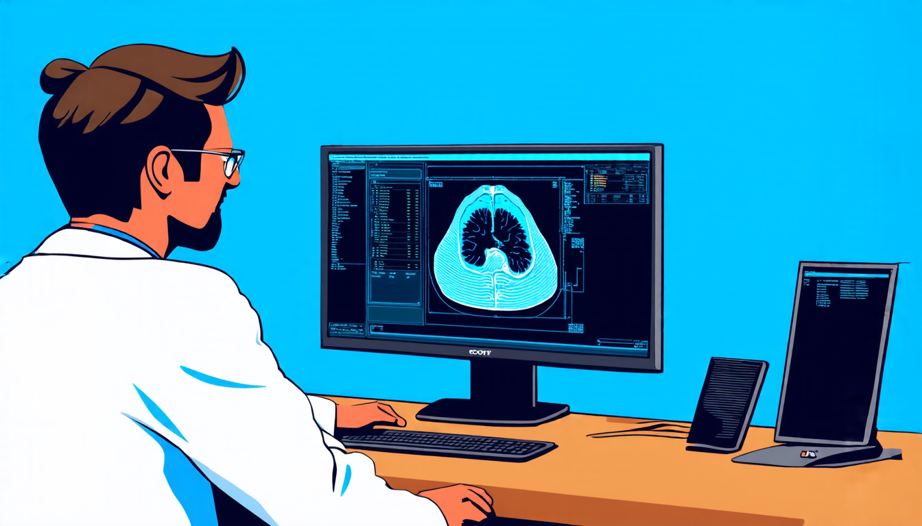
Scientists have made a significant breakthrough in the field of medical imaging, developing a new method for reconstructing computed tomography (CT) scans using artificial intelligence. This innovative approach could revolutionize the way doctors diagnose and treat patients.
The current CT scanning process involves taking X-ray images from multiple angles and then reconstructing them into a detailed 3D image of the body. However, this process can be time-consuming and may not always produce accurate results, particularly in cases where the patient has a low dose of radiation or the scan is incomplete.
To address these limitations, researchers have turned to artificial intelligence, specifically score-based diffusion models. These models use complex algorithms to generate images that are similar to real-world data, but with noise removed and details enhanced.
The new method, known as pseudoinverse diffusion, uses a forward stochastic process to simulate the CT scanning process and then reverses it using a score-matching neural network. This approach allows researchers to maintain the noise correlations and noise magnitude of the original scan, resulting in more accurate and detailed images.
One of the key advantages of this method is its ability to reduce noise and artifacts while preserving important details. This is particularly useful in cases where the patient has a low dose of radiation or the scan is incomplete, as it can help doctors make more accurate diagnoses.
The researchers tested their new method using a dataset from The Cancer Genome Atlas Liver Hepatocellular Carcinoma (TCGA-LIHC). They simulated low-dose CT scans and then used the pseudoinverse diffusion model to reconstruct them. The results showed significant improvements in image quality, with reduced noise and artifacts compared to traditional methods.
The implications of this breakthrough are far-reaching. It could enable doctors to diagnose diseases more accurately and make better treatment decisions, particularly for patients who have undergone low-dose radiation therapy or have incomplete scans.
In addition, the pseudoinverse diffusion model has the potential to be applied to other medical imaging modalities, such as magnetic resonance imaging (MRI) and positron emission tomography (PET). This could lead to a more comprehensive understanding of the human body and improved patient care.
Overall, the development of pseudoinverse diffusion is an exciting advancement in the field of medical imaging. It has the potential to revolutionize the way doctors diagnose and treat patients, and could have significant implications for the future of healthcare.
Cite this article: “Revolutionizing Medical Imaging with Artificial Intelligence”, The Science Archive, 2025.
Medical Imaging, Artificial Intelligence, Ct Scans, Computed Tomography, Score-Based Diffusion Models, Pseudoinverse Diffusion, Neural Networks, Image Reconstruction, Medical Diagnosis, Healthcare Innovation