
Sunday 30 March 2025
Researchers have made a significant breakthrough in understanding the intricate relationships between muscle fibers, nerve endings, and electrical activity in the body. By combining advanced imaging techniques with sophisticated computational methods, scientists have been able to reconstruct three-dimensional models of muscle tissue and analyze the spatial distribution of neuromuscular junctions (NMJs) within them.
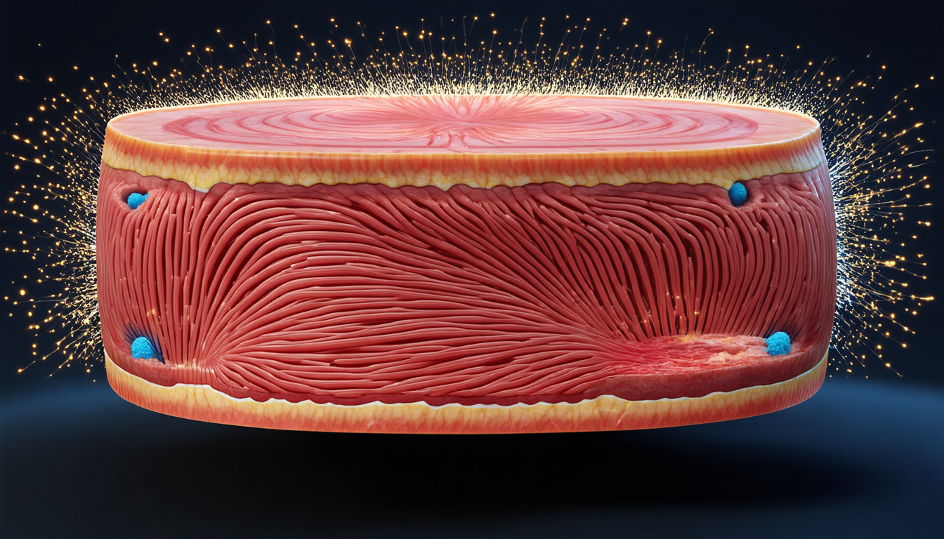
The study focused on the rat soleus muscle, a common model for understanding human muscle function and disease. The researchers used histological slides, which are thin slices of tissue stained with special dyes to highlight specific features, to reconstruct the three-dimensional structure of the muscle. They then extracted the coordinates of NMJs from these images and analyzed them using various methods.
One approach involved dividing the muscle into a grid of small cubes, or voxels, and counting the number of NMJs within each voxel. This allowed the researchers to visualize the density of NMJs throughout the muscle and identify regions with higher concentrations of these critical connections.
Another method used clustering algorithms to group NMJs based on their proximity to one another. This revealed clusters of NMJs that were more densely packed than others, suggesting that these areas may play a key role in coordinating muscle activity.
The researchers also overlaid electrical activity recorded from electrodes placed on the surface of the muscle onto the three-dimensional model. This allowed them to visualize how the electrical signals generated by the muscle fibers interacted with the NMJs and other structures within the tissue.
One of the most striking findings was the correlation between the distance of an NMJ from an electrode and the strength of the electrical signal recorded at that location. The researchers found that as the distance between an NMJ and an electrode increased, the signal strength decreased, indicating that the signals were being attenuated by the tissue.
These results have significant implications for our understanding of muscle function and disease. By better understanding how NMJs are distributed throughout the muscle and how they interact with electrical activity, researchers may be able to develop new treatments for conditions such as muscular dystrophy and amyotrophic lateral sclerosis (ALS).
The study’s findings also highlight the potential of advanced imaging techniques and computational methods in biomedical research. By combining these approaches, scientists can gain a deeper understanding of complex biological systems and develop new insights into human health and disease.
In addition to their scientific significance, the study’s results may have practical applications in the development of prosthetic limbs and other medical devices.
Cite this article: “Unraveling the Complex Interplay Between Muscle Fibers and Nerve Endings”, The Science Archive, 2025.
Muscle Fibers, Nerve Endings, Electrical Activity, Biomedical Research, Advanced Imaging Techniques, Computational Methods, Neuromuscular Junctions, Muscle Tissue, Rat Soleus Muscle, Human Disease