Sunday 30 March 2025
Deep learning and classical computer vision techniques have been combined in a new study that has achieved impressive results in medical image analysis. The research team, comprising of experts from various institutions, has demonstrated the strengths of both approaches by applying them to different imaging modalities.
Medical imaging plays a crucial role in understanding body structures and aiding diagnosis. However, analyzing images can be a complex task, especially when dealing with multiple imaging modalities such as magnetic resonance imaging (MRI), computed tomography (CT), and dermoscopy. To tackle this challenge, the researchers employed a combination of deep learning and classical computer vision techniques.
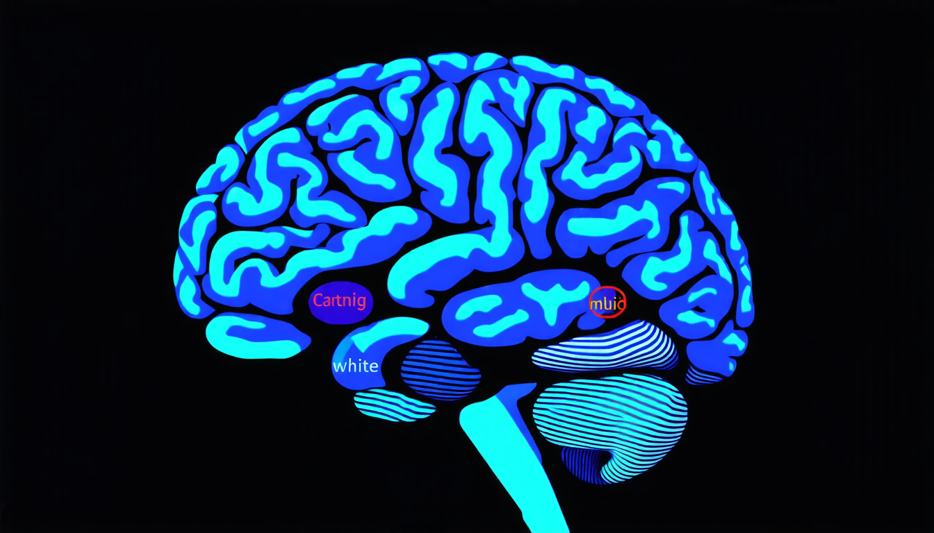
The study focused on three specific tasks: brain MRI tissue segmentation, lung CT image registration, and skin lesion classification from dermoscopic images. Brain tissue segmentation involves identifying different types of tissues in an MRI scan, which is essential for understanding neurological disorders such as Alzheimer’s disease. Lung CT image registration is crucial for monitoring changes in lung function over time in patients with chronic obstructive pulmonary disease (COPD). Skin lesion classification aims to identify skin cancers from images taken using dermoscopy.
The researchers used a range of techniques, including convolutional neural networks (CNNs), residual networks, and multi-atlas methods. CNNs are particularly well-suited for image analysis tasks due to their ability to learn features automatically. Residual networks, on the other hand, are designed to handle large datasets and can be trained using massive amounts of data.
The results of the study show that the combination of deep learning and classical computer vision techniques outperformed traditional methods in many cases. For brain MRI tissue segmentation, the CNN-based approach achieved a Dice coefficient of 0.9397, which is a measure of overlap between predicted and actual segmentations. In contrast, multi-atlas methods resulted in a lower Dice coefficient of 0.7267.
For lung CT image registration, the researchers found that classical Elastix-based methods outperformed deep learning models, achieving a minimum target registration error (TRE) of 6.68 mm. However, high-resolution ResNet models performed competitively with TREs of up to 7.40 mm.
In skin lesion classification, ensembles of deep learning models such as InceptionResNetV2 and ResNet50 excelled, achieving accuracies of up to 93.62% in multiclass classification tasks.
Cite this article: “Combining Deep Learning and Classical Computer Vision Techniques for Medical Image Analysis”, The Science Archive, 2025.
Medical Image Analysis, Deep Learning, Computer Vision, Mri, Ct Scan, Dermoscopy, Brain Tissue Segmentation, Lung Ct Image Registration, Skin Lesion Classification, Convolutional Neural Networks


