

Sunday 06 April 2025
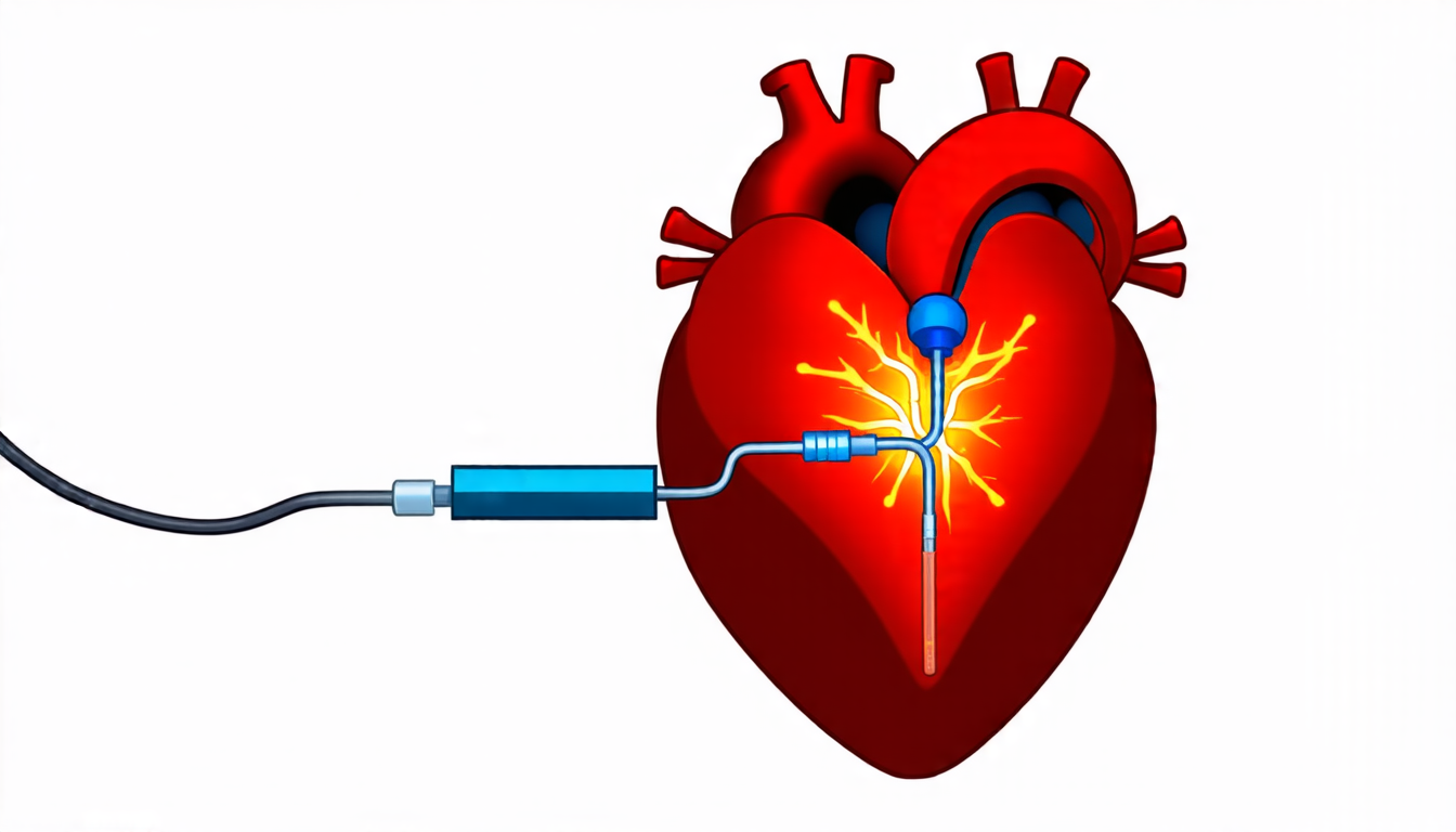
A team of researchers has developed a new approach to detecting and tracking catheters in X-ray fluoroscopy images, which could improve image guidance during minimally invasive heart surgeries.
The traditional method for detecting catheters in these images involves manual annotation by clinicians, which is time-consuming and prone to errors. Automated detection methods have been proposed, but they often require large amounts of labeled training data and can be sensitive to variations in image quality and patient anatomy.
The researchers took a different approach, using a convolutional neural network (CNN) trained on a combination of publicly available datasets and privately collected images from cardiac catheterization procedures. The CNN was designed to detect not only the catheters themselves but also their electrodes, which are critical for ensuring accurate navigation during the procedure.
To improve performance, the team introduced a multi-task learning strategy that allowed the network to learn from both detection and segmentation tasks simultaneously. This approach helped the model develop a more robust understanding of the images and improved its ability to generalize to new data.
The researchers also developed a novel method for dynamically adjusting sample weights during training, which allowed them to prioritize more challenging samples and improve overall performance. This technique could be applied to other medical imaging tasks where there is an imbalance in the distribution of training data.
In addition to improving detection accuracy, the new approach was shown to reduce processing time compared to traditional methods. This is critical for real-time image guidance during procedures, where delays can have serious consequences.
The potential benefits of this technology are significant. Improved catheter detection and tracking could enhance patient safety, reduce procedure times, and enable more complex procedures to be performed. Additionally, the multi-task learning approach could be adapted to other medical imaging tasks, such as tumor segmentation or fracture detection.
While there is still much work to be done before this technology can be deployed in clinical settings, the researchers’ results are an important step forward in developing more accurate and efficient methods for detecting and tracking catheters in X-ray fluoroscopy images.
Cite this article: “Real-Time Catheter Detection and Segmentation in X-Ray Images via Multi-Task Learning: A Breakthrough in Interventional Cardiology Imaging”, The Science Archive, 2025.
Medical Imaging, Catheter Detection, Convolutional Neural Network, Multi-Task Learning, X-Ray Fluoroscopy, Image Guidance, Cardiac Catheterization, Deep Learning, Medical Robotics, Computer-Assisted Surgery





