Monday 07 April 2025
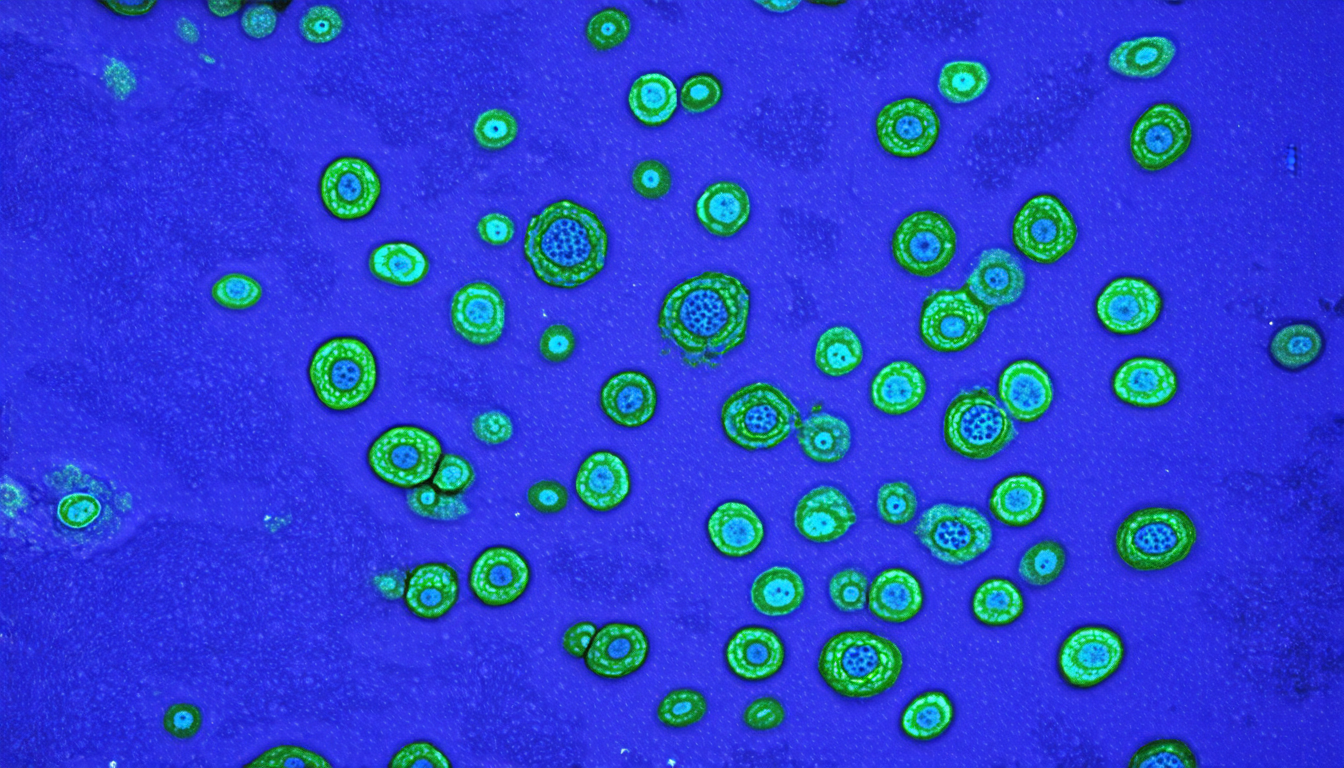
A team of researchers has developed a new approach to detecting nuclei in histopathology whole slide images, which could revolutionize the way we diagnose and treat diseases.
The process of diagnosing cancer typically involves examining tissue samples under a microscope to look for abnormalities such as irregularly shaped cells or clusters of cells. However, this can be a time-consuming and labor-intensive process, requiring experts to manually examine large numbers of slides. The development of machine learning algorithms has the potential to automate this process, allowing for faster and more accurate diagnoses.
One challenge in developing these algorithms is that traditional methods rely on detecting nuclei in individual patches of tissue, without taking into account the larger context of the surrounding tissue. This can lead to inaccuracies and a lack of precision in the detection of abnormalities.
The new approach developed by the researchers addresses this issue by aggregating contextual information from previously visited sliding windows during whole-slide inference. This allows the algorithm to take into account not just individual patches of tissue, but also the larger patterns and structures present in the surrounding tissue.
The researchers used a combination of convolutional neural networks (CNNs) and transformers to develop their algorithm. The CNNs were used to extract features from individual patches of tissue, while the transformers were used to aggregate contextual information from previously visited sliding windows.
In testing their algorithm, the researchers found that it was able to achieve significantly better results than traditional methods, with a precision rate of 91.08% compared to 66.21% for traditional methods. The algorithm also ran much faster, taking an average of 109.98 seconds per whole slide image compared to 356.87 seconds for traditional methods.
The potential applications of this technology are vast. For example, it could be used to develop more accurate and efficient systems for diagnosing cancer, or to help researchers identify new biomarkers for disease detection. It could also be used in other fields such as dermatology, where the ability to quickly and accurately detect skin lesions could revolutionize the way we diagnose and treat conditions like melanoma.
The development of this technology is a testament to the power of machine learning and its potential to transform our understanding of the world around us. By combining cutting-edge algorithms with high-performance computing, researchers are able to develop solutions that were previously unimaginable.
Cite this article: “Context-Aware Nucleus Detection in Histopathology Images via Multi-Scale Feature Fusion and Attention Mechanism”, The Science Archive, 2025.
Histopathology, Machine Learning, Nuclei Detection, Cancer Diagnosis, Whole Slide Images, Convolutional Neural Networks, Transformers, Precision Medicine, Medical Imaging, Biomarkers.


