Wednesday 16 April 2025
The quest for high-precision medical image segmentation has long been a challenge in the field of computer vision. Researchers have employed various techniques, from convolutional neural networks (CNNs) to transformers, but the results often fall short. Recently, a team of scientists introduced Deconver, a novel network that leverages nonnegative deconvolution as a learnable module within a U-shaped architecture.
Deconver’s design is centered around the idea of replacing computationally expensive attention mechanisms with efficient deconvolution operations. This approach enables the network to effectively restore high-frequency details while suppressing artifacts. The result is a model that consistently outperforms leading baselines in both 2D and 3D medical image segmentation tasks.
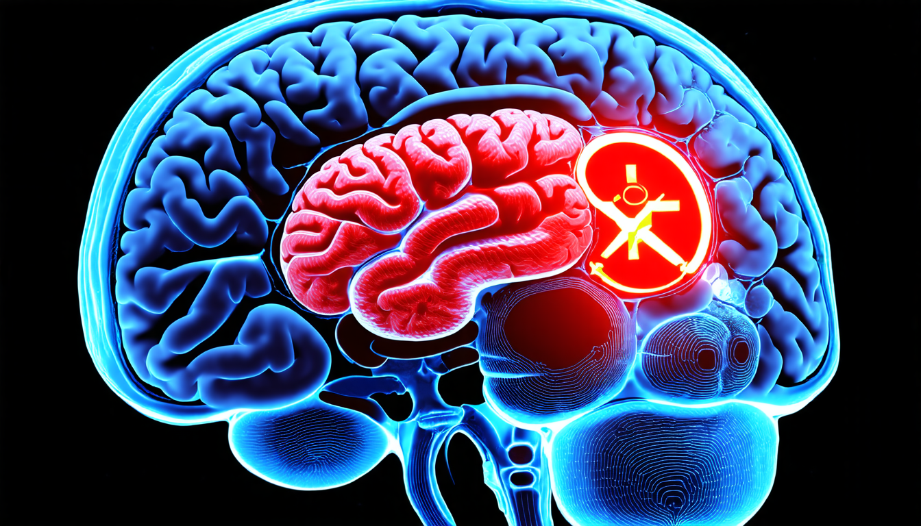
The team tested Deconver on four diverse datasets, including ISLES’22, BraTS’23, GlaS, and FIVES. In each case, Deconver demonstrated impressive results, achieving state-of-the-art performance while significantly reducing computational costs. For instance, in the 3D brain tumor segmentation task, Deconver reduced the number of floating-point operations (FLOPs) by up to 90% compared to leading attention-based models.
Deconver’s architecture is built upon a U-shaped design, which combines features from both CNNs and transformers. The network consists of two main components: a encoder that extracts contextual information and a decoder that generates the final segmentation mask. The key innovation lies in the use of nonnegative deconvolution as a learnable module within the decoder.
Nonnegative deconvolution is a technique borrowed from image restoration, where it’s used to separate signals from noise. In Deconver, this approach enables the network to effectively restore high-frequency details while suppressing artifacts. The result is a model that produces highly accurate segmentation masks with minimal computational overhead.
One of the most impressive aspects of Deconver is its ability to generalize across different datasets and tasks. The team demonstrated that the network could be fine-tuned for specific applications, such as brain tumor segmentation or fundus vessel segmentation, without sacrificing performance.
Deconver’s potential impact on medical imaging is vast. With its ability to produce highly accurate segmentation masks with minimal computational overhead, this network could revolutionize clinical workflows and enable real-time decision-making in a variety of medical applications.
Cite this article: “Revolutionizing Medical Image Segmentation with Deconver: A Deep Learning Approach”, The Science Archive, 2025.
Medical Image Segmentation, Deconver, Nonnegative Deconvolution, U-Shape Architecture, Cnns, Transformers, Attention Mechanisms, 2D Medical Images, 3D Medical Images, Brain Tumor Segmentation, Fundus Vessel Segmentation


