Sunday 09 March 2025
Deep learning has revolutionized many fields, including medical imaging, where it’s used to diagnose and treat a wide range of diseases. One of the key challenges in medical imaging is segmenting objects from images, such as tumors or organs, which requires developing accurate models that can distinguish between different structures.
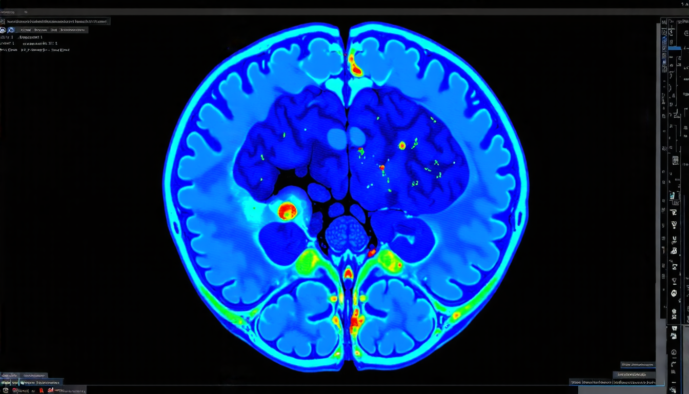
A team of researchers has proposed a new approach to tackling this problem by using distance maps, which are visual representations of the distance between each pixel in an image and the closest boundary of the object being segmented. By incorporating these distance maps into their model, the researchers were able to improve the accuracy of tumor segmentation in medical images.
The team used a combination of two different models: one for classification-based segmentation, which is commonly used in deep learning, and another for regression-based segmentation, which is less common but has been shown to be effective in certain applications. The classification model uses a traditional convolutional neural network (CNN) architecture to predict the probability map of the object being segmented, while the regression model uses a different CNN architecture to predict the distance map.
The key innovation of this approach is the way that the two models are combined. The classification model provides an initial estimate of the object’s boundary, which is then used as input to the regression model. This allows the regression model to refine its predictions based on the information provided by the classification model, leading to more accurate results.
The researchers tested their approach on a dataset of computed tomography (CT) scans and magnetic resonance imaging (MRI) scans, and found that it outperformed state-of-the-art methods in terms of accuracy. The approach was also able to handle images with varying levels of noise and artifacts, which is an important consideration in medical imaging.
The use of distance maps as a regularization term has several benefits. It allows the model to learn more robust features that are less sensitive to noise and artifacts, and it provides a way to incorporate prior knowledge about the object being segmented. For example, if the object is known to have a certain shape or texture, this information can be incorporated into the distance map to guide the segmentation process.
The researchers also experimented with different types of distance maps, including norm inverse distance maps (NI-DMs) and sign norm inverse distance maps (SNI-DMs). They found that NI-DMs performed best overall, but SNI-DMs were effective in certain situations.
Cite this article: “Distance Maps Improve Tumor Segmentation Accuracy in Medical Imaging”, The Science Archive, 2025.
Medical Imaging, Deep Learning, Tumor Segmentation, Distance Maps, Convolutional Neural Networks, Regression-Based Segmentation, Classification-Based Segmentation, Computed Tomography, Magnetic Resonance Imaging, Norm Inverse Distance Maps


