

Monday 31 March 2025
Deep learning has revolutionized many fields, from image recognition to natural language processing. But what happens when you combine these two areas and apply them to medical imaging? A team of researchers has made significant strides in this area by developing a new model that can not only recognize images but also generate reports based on those images.
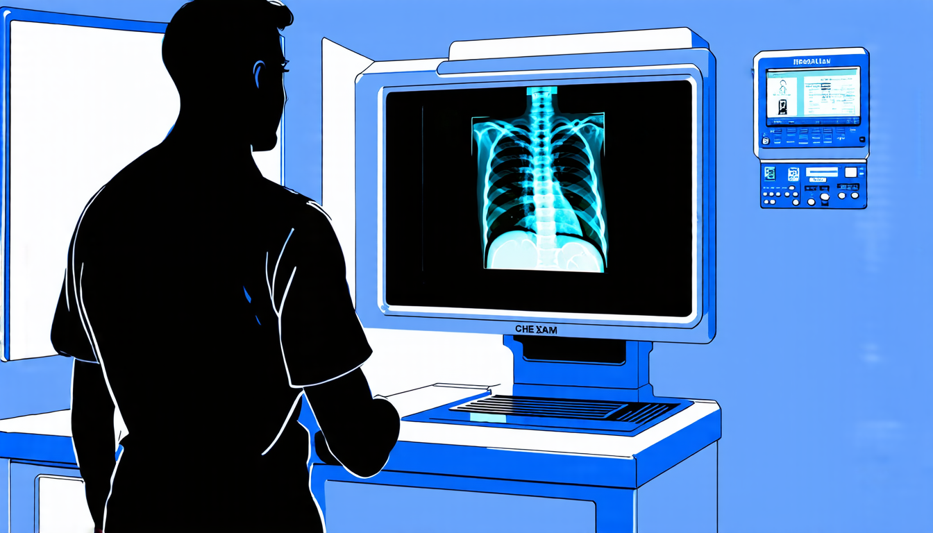
The new model, called CoCa- CXR, is designed specifically for chest X-ray (CXR) images. These images are used to diagnose a wide range of respiratory and cardiovascular conditions, from pneumonia to heart failure. Traditionally, radiologists have interpreted these images by hand, but this can be time-consuming and prone to errors.
CoCa-CXR uses a deep learning algorithm to analyze CXR images and generate reports that are comparable in quality to those written by human radiologists. The model is trained on a large dataset of CXR images and corresponding reports, which allows it to learn patterns and relationships between the two.
One of the key features of CoCa-CXR is its ability to recognize temporal changes in CXR images over time. This means that the model can detect subtle changes in an image from one exam to the next, such as worsening pneumonia or improved lung function.
The researchers tested CoCa-CXR on a dataset of 20,000 CXR images and found that it performed almost as well as human radiologists in terms of accuracy and completeness. The model was also able to generate reports that were free from errors and consistent with the image findings.
CoCa-CXR has significant implications for healthcare systems around the world. By automating the reporting process, radiologists will be freed up to focus on more complex cases and provide better patient care. Additionally, CoCa-CXR can help reduce costs by reducing the need for manual reporting and improving diagnostic accuracy.
The researchers are now working to fine-tune the model and integrate it into clinical practice. They hope that CoCa-CXR will become a valuable tool for radiologists and clinicians alike, helping to improve patient outcomes and streamline healthcare workflows.
In recent years, deep learning has made significant strides in medical imaging. From detecting diabetic retinopathy to diagnosing breast cancer, AI-powered models have shown impressive results. But CoCa-CXR takes things to the next level by integrating image recognition with natural language processing. This marriage of technologies holds great promise for healthcare, and it will be exciting to see where this technology goes in the future.
The model’s ability to recognize temporal changes is particularly noteworthy.
Cite this article: “Automating Chest X-Ray Reporting with Deep Learning”, The Science Archive, 2025.
Chest X-Ray, Deep Learning, Natural Language Processing, Medical Imaging, Image Recognition, Radiologists, Reports, Accuracy, Completeness, Temporal Changes





